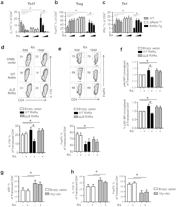
Fig. 6. Gain and loss of function effects of RARα on T cell differentiation and Akt/mTOR activity.
(a-c) Impact of RARα dose and RA (0, 1, 10, 50 nM) on Th17/Treg/Th1 polarization in vitro. (d) Impact of the expression of WT and ΔLB RARα on the polarization of naive ΔRaraLCK CD4 T cells into Th17 cells. (e) Impact on the polarization of naive ΔRaraLCK CD4 T cells into FoxP3+ T cells. (f) Impact on the phosphorylation of Akt (S473) and rS6 (S235/236) in ΔRaraLCK CD4 T cells. (g) Impact of the constitutively active form of Akt (Myr-Akt) on mTOR activation (rS6 phosphorylation). (h) Impact of Myr-Akt on Th17 versus Treg differentiation. For panel d, e, and h, ΔRaraLCK T helper cells were transduced after 16 h culture with anti-CD3/CD28 (5 μg/mL and 2 μg/mL, respectively) with RARα- or Myr-Akt-expressing retrovirus and subsequently cultured in Th17- or Treg-polarizing culture for 3 additional days prior to flow cytometry analysis. For panels f and g, ΔRaraLCK T helper cells were similarly transduced and cultured in Th17-polarizing culture for 20 h prior to flow cytometry analysis. Representative and combined data (n=4–7) from more than 4 independent experiments are shown. *Significant differences (P values <0.05) between indicated groups by repeated measures two-way ANOVA with Bonferroni.

