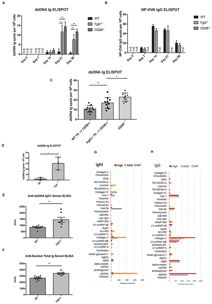
Figure 7.
Fgl2 is important for limiting autoantibodies. (a-b) Wild-type and Fgl2−/− mice were immunized by NP-OVA/CFA (with additional heat killed dried Mycobacterium tuberculosis). Inguinal lymph nodes were harvested at the time points indicated and the cells were subjected to ELISPOT assay to detect dsDNA Ig and NP-OVA IgG ASCs. (c) Sorted 10,000 WT or Fgl2−/− CD19−CD4+CXCR5+PD1+Foxp3+ TFR cells were transferred into CD28−/− mice. The mice were then immunized with NP-OVA/CFA (with additional heat killed dried Mycobacterium tuberculosis) for 21 days and dsDNA Ig ASCs were detected by ELISPOT (d) Splenocytes from 12-month-old wild-type and Fgl2−/− mice were subjected to dsDNA Ig ELISPOT. (e-f) Anti-dsDNA IgG1 and ANA in sera from 12-month-old wild-type and Fgl2−/− mice were measured by ELISA. (g-h) An array of lupus IgM and IgG autoantibodies were measured in sera from 12-month-old wild-type (n = 6) and Fgl2−/− mice (n = 9) using autoantigen microarray. Fgl2−/− mice with mild diseases (n = 5) had either normal histology or mild dermatitis while the one with high, severe diseases (n = 4) had inflammation in multiple organs, including severe dermatitis, reactive changes in the spleen, ileitis, enlarged Peyer’s Patches and increased lymphoid clusters in lung. The results were pooled from 2-3 independent experiments. The autoantigen microarray results for (g-h) are from one independent experiment. Differences between two groups shown were compared using Kruskal-Wallis tests followed by post hoc Dunn’s tests for multiple comparisons for (a) to (c) and two-tailed unpaired Welch’s T tests for (d) to (f) (n.s; * p <0.05, ** p<0.01 and *** p<0.001). All plots show the mean ± SD.

