
Figure 5. αCD28 Ab accelerates proliferation and activation of Foxp3+ Treg in memory septic mice.
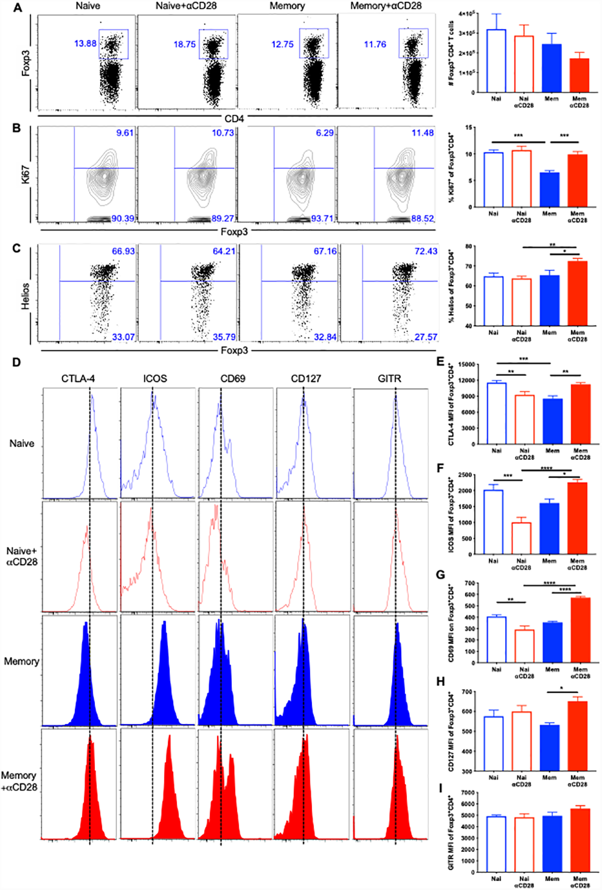
Memory mice and age-matched naïve controls underwent CLP, followed by injection of αCD28 Ab or saline. Mice were sacrificed and spleens were harvested at 24 hours post-CLP. (A) Absolute numbers of Foxp3+ Treg cells among four groups (n=8–9/group). (B) The proliferation of Treg cells was assessed by Ki67 staining. The percentages of Ki67+ Treg cells in the four groups (n=8–9/group) are shown. (C) Representative flow plots and summary data of the percentage of Helios+ in Tregs. (D-I) Flow histograms and summary data of CTLA-4 (E), ICOS (F), CD69(G), CD127 (H) and GITR (I) in memory vs naïve, memory vs memory+αCD28 and naïve vs naïve+αCD28 groups (n=4–9/group). The data were pooled from two independent experiments. Data represented as mean ± SEM and analyzed with one-way ANOVA analysis and Turkey multiple comparison test. *, p<0.05, **, p<0.01, ***, p<0.001 and****, p<0.0001.