
Figure 8. IL-10 blockade induces downregulation of Bcl-xL and increased T cell apoptosis in memory mice treated with αCD28 Ab.
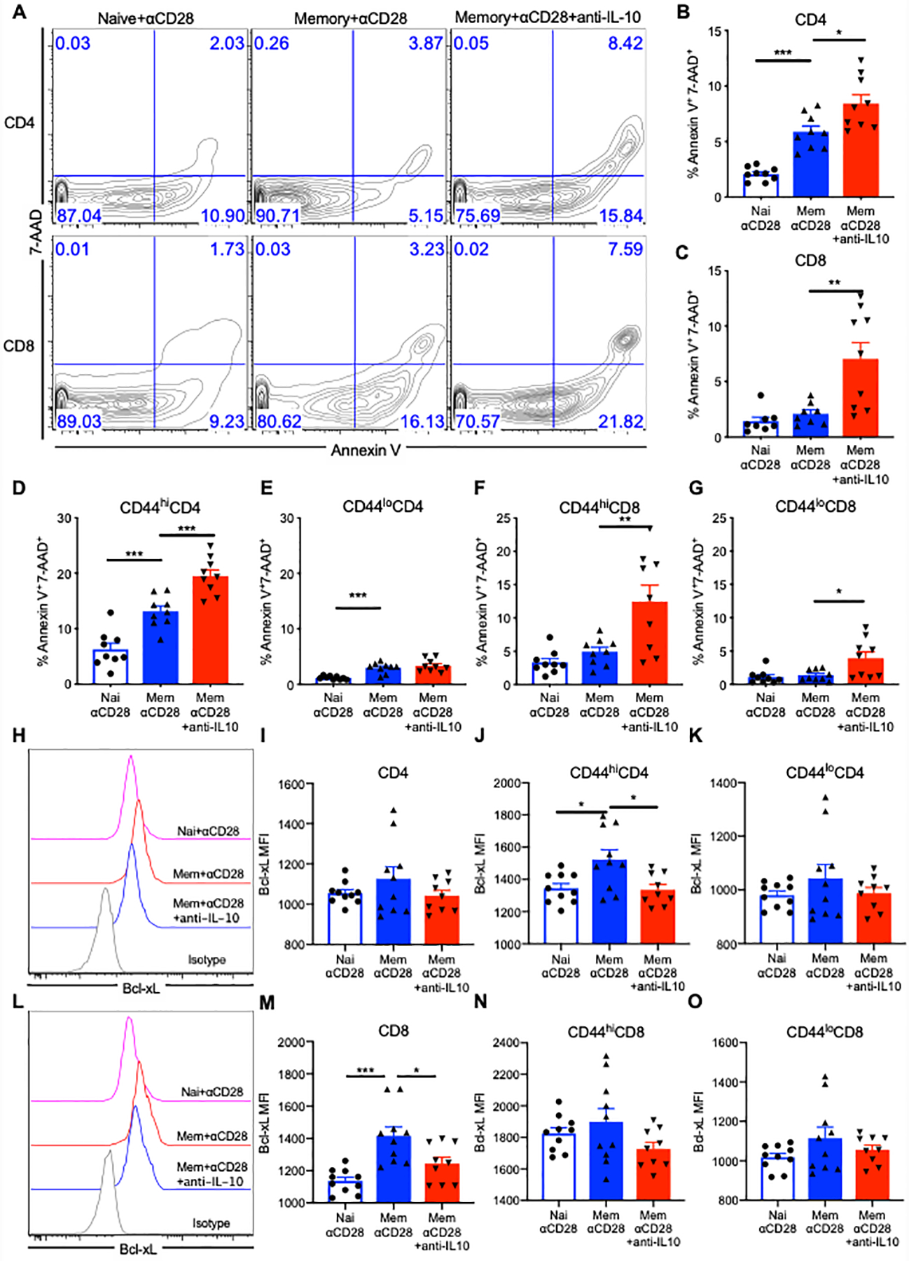
Memory and naïve mice received CLP and αCD28 Ab followed by either anti-IL-10 Ab or isotype IgG Ab. Splenocytes were stained for Annexin V and 7-AAD for apoptosis analysis of T cells and intracellularly stained with anti-Bcl-xL Ab among naïve mice with αCD28 Ab (n=8) vs. memory mice treated with αCD28 Ab (n=9) vs. memory mice treated with αCD28 Ab and anti-IL-10 Ab (n=9) at 24 hours after CLP. (A) Representative flow cytograms indicating the apoptosis of CD4+ and CD8+ T cells among three groups. (B) Summary of percentage of apoptotic CD4+ T cells. (C) Summary of percentage of apoptotic CD8+ T cells. (D and E) Data depicting the frequency of apoptotic memory (CD44hi) and naïve (CD44lo) CD4+ T cells. (F and G) Data depicting the frequency of apoptotic CD44hiCD8+ and CD44loCD8+ T cells. (H and I) Representative flow cytograms and summary data indicating Bcl-xL expression in CD4+ T cells among naïve + αCD28 vs memory + αCD28 vs memory + αCD28 + anti-IL-10 groups. (J and K) Summary of Bcl-xL MFI in CD44hiCD4+ and CD44loCD4+ T cells. (L and M) Representative flow cytograms and summary data indicating Bcl-xL expression in CD4+ T cells among three groups. (N and O) Summary of Bcl-xL MFI in CD8+, CD44hiCD8+ and CD44loCD8+ T cells. The results depict two independent experiments. Data represented mean ± SEM. Groups were compared with one-way ANOVA analysis and Turkey multiple comparison test. *, p<0.05, **, p<0.01 and ***, p<0.001.