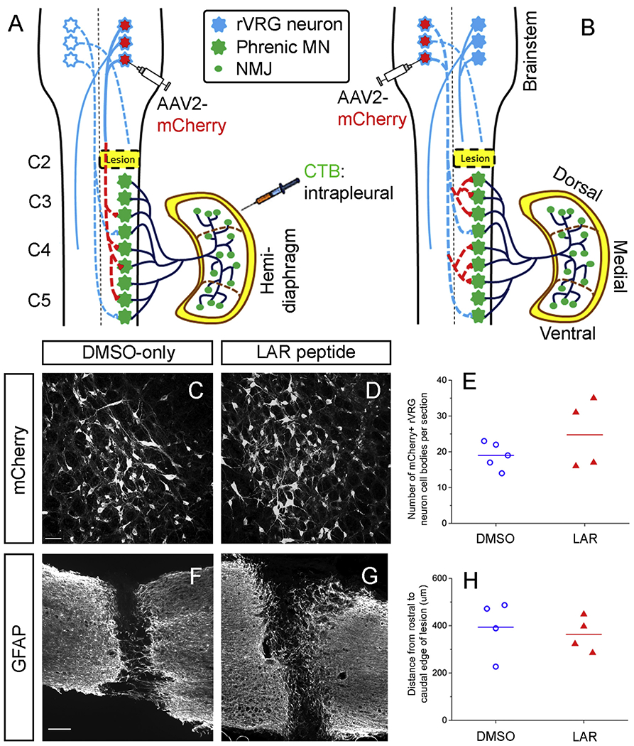
Fig. 4.

Labeling of rVRG-PhMN circuitiy. (A) To determine whether LAR peptide treatment promoted rVRG axon regeneration, we selectively transduced rVRG neurons within the medulla ipsilateral to the hemisection with AAV2-mCherry. (B) In a separate cohort of rats, we examined whether sprouting of spared rVRG axons originating in the contralateral medulla was induced by LAR peptide by injecting the AAV2-mCherry anterograde tracer into the contralateral rVRG and then quantifying sprouting of these axons within the PhMN pool on the side of the hemisection (i.e. contralateral to the location of their cell bodies). In both cohorts, we intrapleurally injected Cholera Toxin B Subunit (CTB) to selectively label PhMN cell bodies within the cervical spinal cord. There was no difference in the number of AAV2-mCherry transduced rVRG neuron cell bodies in the medulla between the DMSO-only (C) and LAR peptide (D) groups (P = 0.32, n = 4 per group); quantification in (E). Sagittal spinal cord sections immunolabeled for GFAP from DMSO-only control (F) and LAR peptide (G) show that LAR peptide did not alter lesion size after C2 hemisection. (H) Quantification of GFAP-labeled sections shows no difference in lesion size as measured by the rostral-caudal distance from the rostral to caudal borders of the lesion (P = 0.67, n = 4 rats per group).
