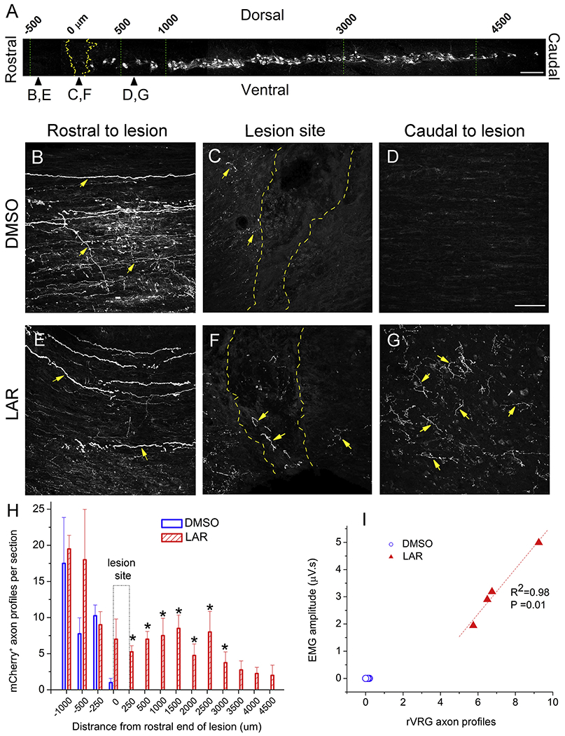
Fig. 5.

LAR peptide promoted rVRG axon regeneration through the lesion and into the intact caudal spinal cord. (A) Sagittal section of cervical spinal cord showing the columnar organization of CTB-labeled PhMNs extending from C3 to C5, the locations (i.e. rostral-caudal distances relative to the hemisection site) of rVRG axon growth analysis, and the location of the representative images in panels B-G; scale bar in (A): 250 μm. Representative images of sagittal sections from DMSO-only or LAR peptide treated rats injected with AAV2-mCherry into the ipsilateral rVRG: (B,E) rostral to lesion site, (C,F) lesion site, (D,G) caudal to lesion site. These images from the two groups were always taken from the same rostral-caudal position/distance relative to the lesion epicenter. Yellow arrows in panels B-G denote mCheriy-labeled rVRG axons. Dotted yellow lines in panels C and F denote the rostral and caudal lesion-intact borders. Scale bar: 100 μm. (H) Quantification shows the numbers of ipsilateral-originating mCheriy-labeled rVRG axons at different locations in DMSO-only and LAR peptide group using the rostral end of the lesion as the rostral-caudal starting point. The indicated differences were compared between DMSO-only and LAR peptide groups at each distance (n = 4 animals per group, *P < 0.05). (I) Pearson Correlation was used to examine the correlation between the degree of rVRG axon regeneration at the C3 level and EMG amplitude in the corresponding ventral diaphragm subregion (correlation coefficient for linear fit, R2 = 0.98, P = 0.01, n = 4 animals per group). (For interpretation of the references to colour in this figure legend, the reader is referred to the web version of this article.)
