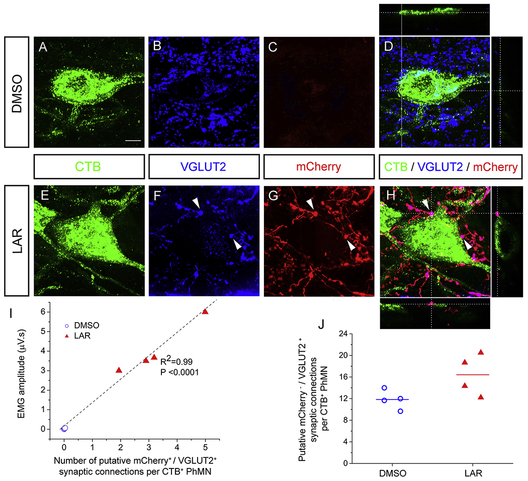
Fig. 6.

LAR peptide promoted putative monosynaptic connection between regenerating rVRG axon and PhMNs. We assessed the number of putative synaptic connections specifically between mCheriy/DsRed-positive rVRG axons and CTB-labeled PhMN cell bodies at level C3 with confocal acquisition of z-stacks and quantification of rVRG axon-PhMN contacts using single-Z section analysis to establish direct apposition of pre-synaptic VGLUT2+/mCheriy+ axon terminals and post-synaptic CTB+ PhMNs. Representative images in panels A-H are compressed z-stacks. (A-D) We observed no putative rVRG-PhMN synapses in the DMSO-only control. (E-H) On the contrary, we observed large numbers of putative rVRG-PhMN synapses in the LAR peptide group. (I) Orthogonal projection shows mCherry+/VGLUT2+ excitatory rVRG axon terminals located directly presynaptic to the soma of a CTB+ PhMN (example putative connections denoted by arrowheads). Scale bar: 100 μm. Quantification of the number of these synaptic connections per individual PhMN at C3 plotted against the EMG amplitude from the corresponding ventral subregion in LAR peptide group (R2 = 0.99; P < 0.0001, n = 4). (J) Quantification of the effects of LAR peptide on putative excitatory synapses from mCherry−/VGLUT2+ non-rVRG inputs per ipsilateral PhMN at C3 (P = 0.07, n = 4 rats per group).
