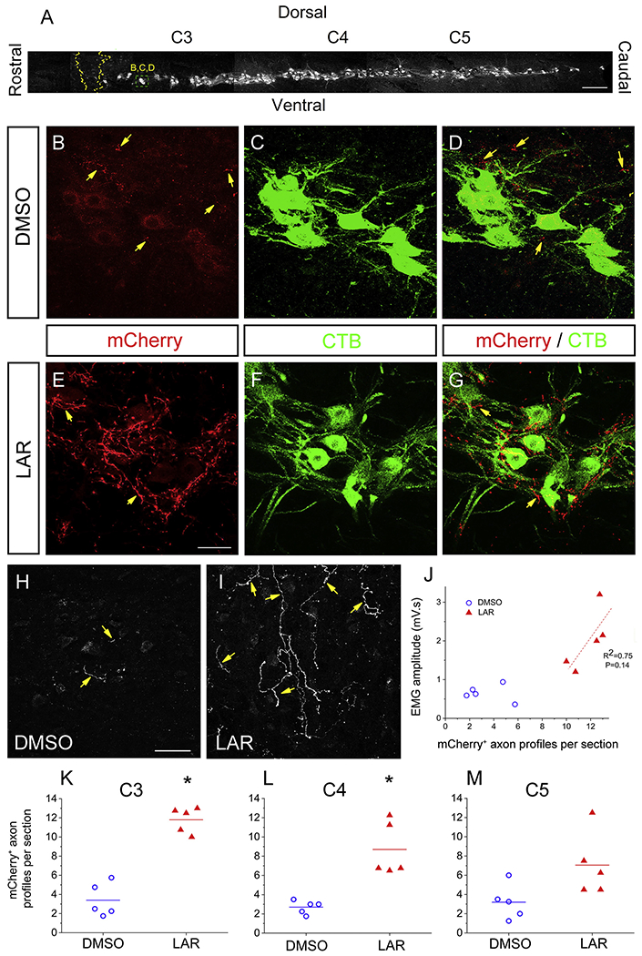
Fig. 7.

LAR peptide promoted sprouting of rVRG axons within the PhMN pool. (A) Sagittal section of cervical spinal cord showing the columnar organization of CTB-labeled PhMNs extending from C3 to C5, the locations of rVRG axon growth analysis, and the location of the representative images in panels B-D as well as E-G. Sagittal spinal cord sections were double-immunolabeled with DsRed (to enhance the mCherry signal) and CTB. We counted numbers of mCherry+ fibers within the phrenic nucleus (i.e. the location of CTB+ PhMNs) at distances of 1.5 mm, 3.0 mm and 4.5 mm caudal to the lesion that correspond to C3, C4 and C5 levels, respectively; scale bar in (A): 250 μm. Representative confocal images show PhMNs labeled with CTB that were surrounded by mCherry+ rVRG axons. Contralateral-originating rVRG axons are present within the PhMN pool on the side of hemisection in both the DMSO-only (B-D) and LAR-peptide treated (E-G) conditions. (H, I) Yellow arrows indicate mCherry-labeled rVRG axons only. Scale bars: 100 μm. (F) The number of these mCherry+ fibers in the LAR peptide group increased significantly compared to DMSO-only control at C3 (E-I, K) and C4 (L), but there was no significant difference at C5 (M) (P < 0.05). (G) Pearson Correlation was used to examine the correlation between the degree of rVRG axon sprouting at the C3 level and EMG amplitude in the corresponding ventral diaphragm subregion in the LAR peptide group (correlation coefficient for linear fit, R2 = 0.75; P = 0.14, n = 5). (For interpretation of the references to colour in this figure legend, the reader is referred to the web version of this article.)
