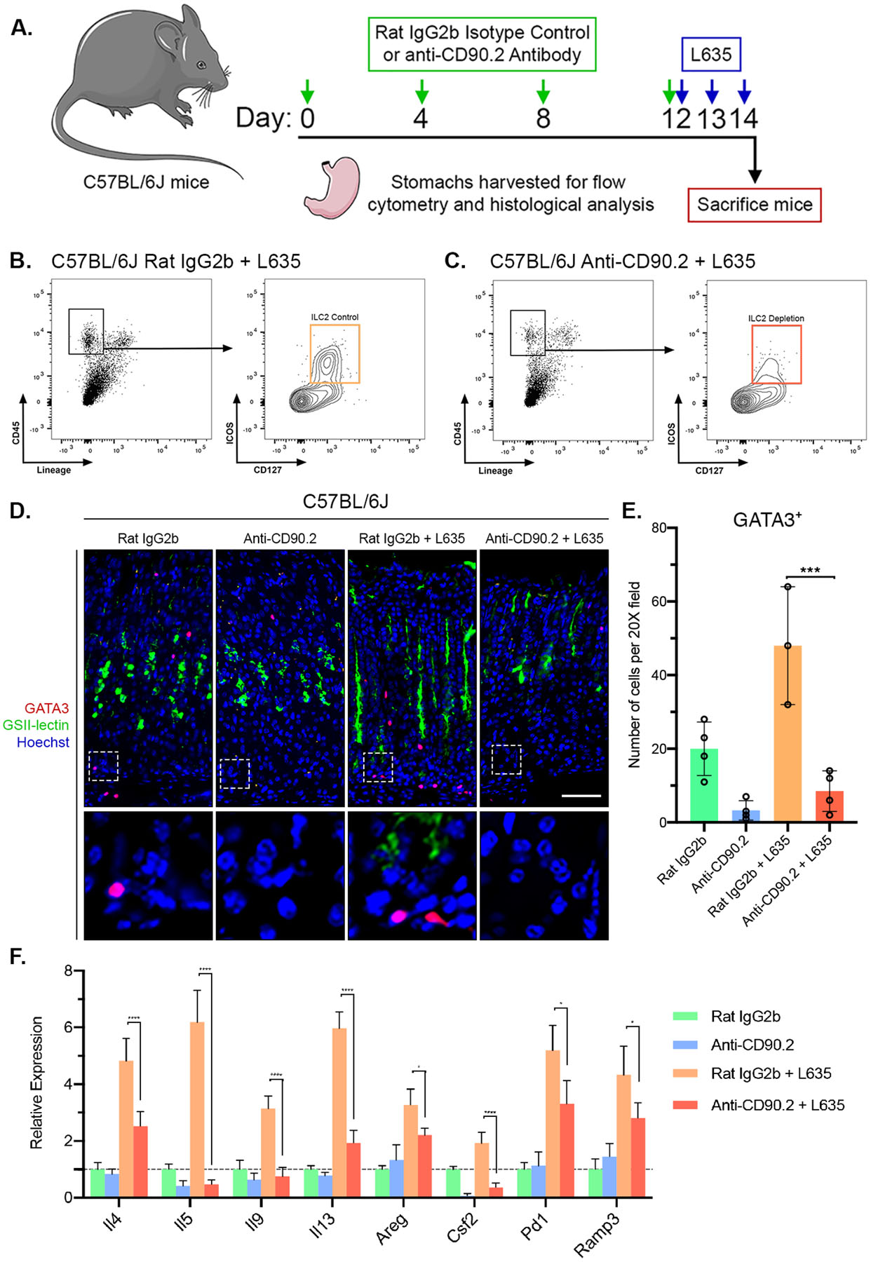
Figure 3. Anti-CD90.2 treatment effectively depletes ILC2s and reduces expression of ILC2-related genes.

(A) Diagram of ILC2 depletion and drug treatments. Rat IgG2b isotype control antibody or Anti-CD90.2 antibody was administered intraperitoneally to wild-type C57BL/6J mice every fourth day for 12 days. Following the final antibody administration, mice were treated with the parietal cell toxic drug L635 by oral gavage daily for three days. Mice were sacrificed 2 hours after final dose of L635, and stomach tissue from Rat IgG2b only mice (n=4), Anti-CD90.2 only mice (n=4), Rat IgG2b + L635 mice (n=3), and Anti-CD90.2 + L635 mice (n=4) was harvested for flow cytometry or histological analysis. Flow cytometric analysis of isolated single cells from (B) Rat IgG2b + L635 and (C) Anti-CD90.2 + L635 stomach tissue. Depletion of CD45+Lin−CD127+ICOS+ ILC2 population with Anti-CD90.2 treatment. (D) Representative images of immunostaining for GATA3 (red), mucin-6 containing granule marker GSII-lectin (green), with nuclear counter stain Hoechst (blue) (scale bars = 100 μm). Magnified inset of gland base (bottom). (E) Quantification of GATA3-positive ILC2s per 20X objective field. (F) Relative mRNA expression of ILC2 related genes (Il4, Il5, Il9, Il13, Areg, Csf2, Pd1, and Ramp3) in each group. Expression values normalized to Rat IgG2b only group. Statistical significance determined by one-way ANOVA with Bonferroni’s post-hoc multiple comparisons test. * for p < 0.05, *** for p < 0.001, and **** for p < 0.0001. Error bars represent mean ± SD.
