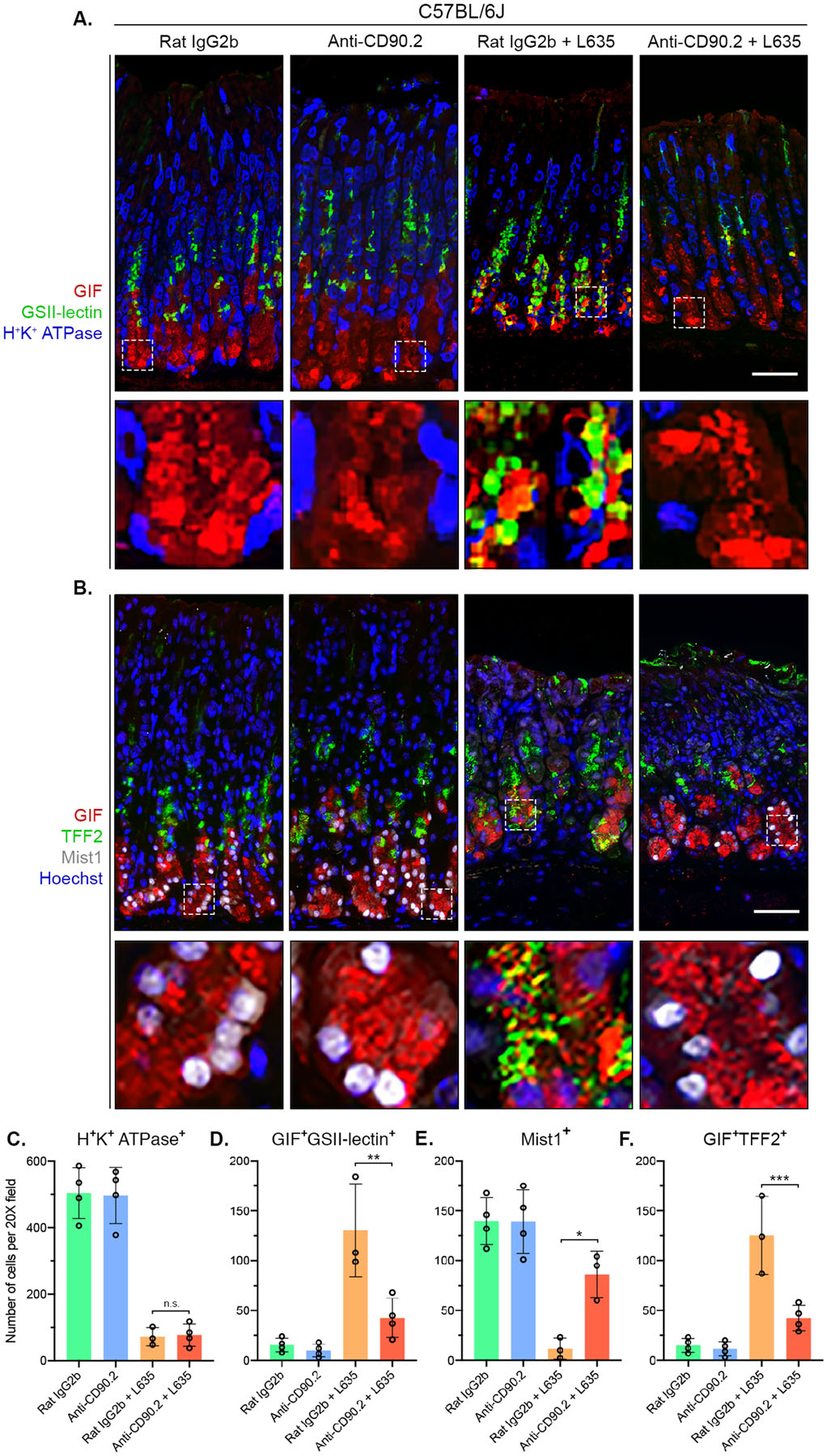
Figure 4. ILC2 depletion blocks chief cell reprogramming to mucin-secreting SPEM after L635-induced injury.

Immunostained sections from Rat IgG2b (n=4), Anti-CD90.2 (n=4), Rat IgG2b + L635 (n=3), and Anti-CD90.2 + L635 (n=4) treated C57BL/6J mice. (A) Representative images of zymogenic granule marker GIF (red), mucin-6 containing granule marker GSII-lectin (green), and parietal cell marker H+/K+ ATPase (blue) (scale bars = 100 μm). Magnified inset of chief cell region (bottom). (B) Representative images of zymogenic granule marker GIF (red), mucin granule marker TFF2 (green), chief cell transcription factor Mist1 (white) with nuclear counter stain Hoechst (blue) (scale bars = 100 μm). Magnified inset of chief cell region (bottom). Quantification of (C) H+K+ ATPase-positive parietal cells (D) GIF and GSII-lectin dual-positive (SPEM) cells (E) Mist1-positive cells and (F) GIF and TFF2 dual-positive (SPEM) cells per 20X objective field. Statistical significance determined by one-way ANOVA with Bonferroni’s post-hoc multiple comparisons test. N.S. for not significant, * for p < 0.05, ** for p < 0.01, and *** for p < 0.001. Error bars represent mean ± SD.
