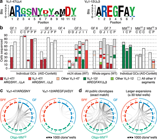
Fig. 4 |. Prominent public clonotypes in gaGCs of GF and Oligo-MM12-colonized mice.
(a) CDRH3 aa sequence logos for B cells bearing public VH1–47/JH4 or JH2 rearrangements of 11 or 12 aa length (left) or VH1–12/JH3, 7 aa length (right), sequenced from GF gaGCs. (b) frequency across mice/samples of B cells fitting public clonotype criteria or carrying other VH1–47 or VH1–12 rearrangements. Each bar represents one sample. Data are for 7 GF mice (GF1–7) and 3 Oligo-MM12-colonized mice (MM121–3). D, duodenal; I, ileal; C, cecal colonic mLN; P, Peyer’s patch. (c) Circos plots showing distribution of VH1–47 and VH1–12 public clonotypes in a second cohort of sex and age-matched SPF, GF, and Oligo-MM12-colonized mice. Each segment represents pooled mLN and PP GC B cells from one mouse, with clones ordered clockwise from largest to smallest. Only clones containing identical CDRH3s are linked (see Online Methods for full description). Gated as in Extended Data Fig. 9b; all samples were sequenced in a single experiment. (d) Circos plot as in (c) showing all public IgH clonotypes shared across mice housed under the indicated conditions. Lines connect clonotypes with the same VH/CDRH3/JH aa sequence. Left, all clones; right, only clones spanning ≥ 30 total wells. In (c, d), only clones found in at least 2 wells from the same mouse were linked.

