Figure 6.
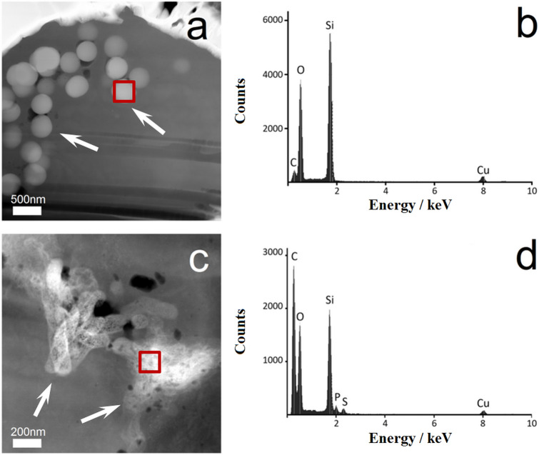
Representative TEM/EDX analyses of FIB-prepared cell lamellae from single NR8383 cells after 24 h of exposure to 100 µg mL−1 unfunctionalized silica particles. The thickness of each TEM lamella was 100 nm. TEM images of cell lamellae were prepared after incubation with microspheres (a) or microrods (c); white arrows indicate particles inside the cells. Corresponding EDX spectra (b and d) of the elements detected within the regions of interest of the cell lamellae (red boxes in a and c) confirmed the presence of silica.

