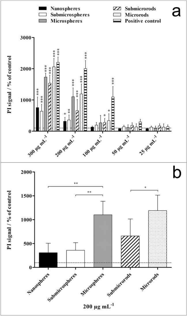
Figure 8.

Viability of NR8383 alveolar macrophages after 16 h incubation with different unfunctionalized silica particles. The cell viability was determined by propidium iodide (PI) staining of non-viable cells and mean PI fluorescence intensity assessment by flow cytometry. Commercial silica particles served as positive control. (a) Comparison of the effects of different particle concentrations and morphologies on the cell viability. (b) Comparison of the effects of different particle morphologies on the cell viability at a particle concentration of 200 µg mL−1. Data are expressed as mean ± SD (n = 3), given as percentage of the control (100%, untreated cells). Asterisks (*) indicate significant differences in comparison to the control (**p < 0.01, ***p < 0.001).
