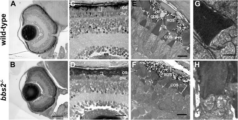
FIGURE 3.
bbs2 – /– mutant zebrafish have shortened and disorganized photoreceptor outer segments at 5 dpf. Representative images of wild-type and bbs2– /– mutants at 5 dpf. (A,B) Semi-thin histological sections show that bbs2– /– mutants have normal retinal lamination. (C,D) Higher magnification images of the dorsal retina show that bbs2– /– mutants have shorter photoreceptor outer segments (OS; white brackets) but gross anatomical structure remains normal. (E–H) Transmission electron microscopy shows shorter and disorganized cone outer segments (COS) in bbs2– /– mutants (white bracket). Cilia architecture appears normal. Scale bar: 50 μm (A,B); 10 μm (C,D); 5 μm (E,F); 1 μm (G,H). COS, outer segments; ROS, rod outer segments.

