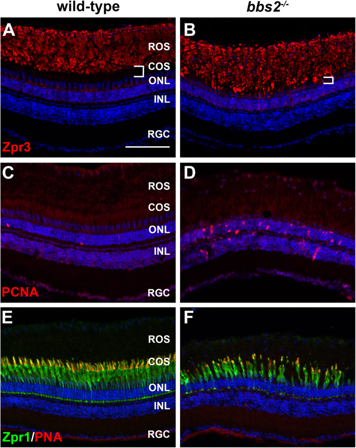
FIGURE 4.
Photoreceptor degeneration in zebrafish retina lacking Bbs2. (A,B) Transverse cryosections of 7 mpf wild-type sibling and bbs2–/– mutant retina stained with Zpr3 (red) to label rhodopsin. The region containing cone outer segments (white brackets) is smaller in bbs2–/– mutants. (C,D) Cryosections of wild-type and bbs2–/– mutants stained with PCNA (red) to label proliferating cells. (E,F) Cryosections of wild-type and bbs2–/– mutants stained with PNA (red) and Zpr1 (green) to label cone outer segments and cone inner segments, respectively. Sections were counterstained with DAPI (blue). Scale bar: 100 μm. RGC, retinal ganglion cells; INL, inner nuclear layer; ONL, outer nuclear layer; COS, cone outer segment layer; ROS, rod outer segment layer.

