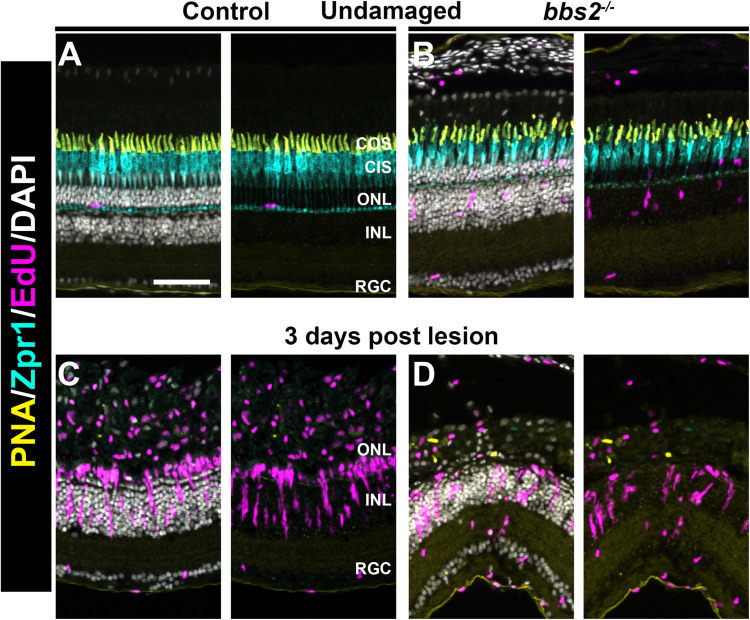
FIGURE 7.
Light damage triggers proliferation of Müller glia in wild-type and bbs2– /– mutants. Transverse cryosections of undamaged retinas from control (A) and bbs2– /– mutant (B) adult (5 mpf). Cryosections were stained with PNA to label cone outer segments (yellow), Zpr1 to stain cone inner segments (cyan), EdU to identify cells that underwent proliferation (magenta) and DAPI (grays). Right panels omit DAPI channel to permit better visualization of EdU labeling. Cone photoreceptors are disorganized and a small number of proliferating cells are seen in the ONL and INL of bbs2– /– mutants. (C,D) Images of transverse cryosections of light damaged control animals (C) and bbs2– /– mutants (D) at 3 days post light lesion. Light damage has largely eliminated photoreceptors and neurogenic clusters of EdU + cells are seen in both groups. Scale bar: 50 μm. RGC, retinal ganglion cells; INL, inner nuclear layer; ONL, outer nuclear layer; CIS, Cone inner segments; COS, cone outer segment layer.

