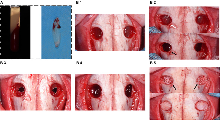
FIGURE 2.
Surgical procedure diagram in a perforated SM model. (A) Preparation of A-PRF. (B1) Symmetrical bone defects were obtained, and bone plates were acquired correspondingly. (B2) SM was detached and elevated from bony walls (marked with white arrow). Rhythmic movement of SM during respiration (black arrow). (B3) SM was perforated with a 1-cm incision (marked with the white dotted box). (B4) CM or A-PRF was placed onto the perforated SM. (B5) The sinuses were filled with DBBM (marked with black arrows). Finally, the bone defects were covered with bone plates.

