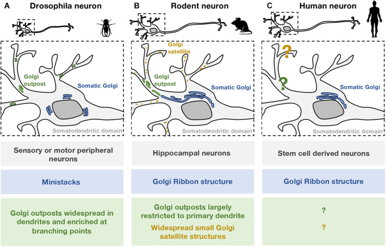
FIGURE 1.
Comparison of somatic and dendritic Golgi structures in different neuronal models. The organization of Golgi structures in (A) Drosophila, (B) rodent, and (C) human neurons are illustrated. Zebrafish neurons are not included as the Golgi structures are not well defined. (A) In Drosophila cell models, most neurons examined are from the peripheral nervous system. In these neurons, the somatic Golgi apparatus (blue) appears as mini-stacks or “ring”-like stacks. GOs (green) are widespread in the dendritic network including the distal dendrites, and are particularly enriched at branching points. Both single- and multi-compartmented Golgi outposts are present in the Drosophila dendritic network. (B) In rodent models, most neurons examined are cultured embryonic hippocampal neurons. In these neurons, the somatic Golgi apparatus is a Golgi ribbon (blue), and appears to extend into the primary dendrite. Stacked GOs (green) are largely restricted to one primary dendrite and are often found in the proximal region. Smaller, non-stacked Golgi satellite structures (orange) are identified in the dendritic arborisation of rodent neurons. (C) In human neurons, the arrangement of secretory organelles, including the Golgi apparatus, is not well defined. A dendritic Golgi in human neurons has yet to be identified. Given the structural differences observed in Drosophila (A) and rodent (B) neurons, conclusions about human neurons should be drawn carefully especially in relation to “Golgi outposts.”

