Figure 3.

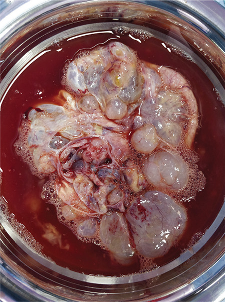
Cut specimen of left ovarian mass showing both solid and cystic areas containing clear fluid and a solid white-colored area

Cut specimen of left ovarian mass showing both solid and cystic areas containing clear fluid and a solid white-colored area