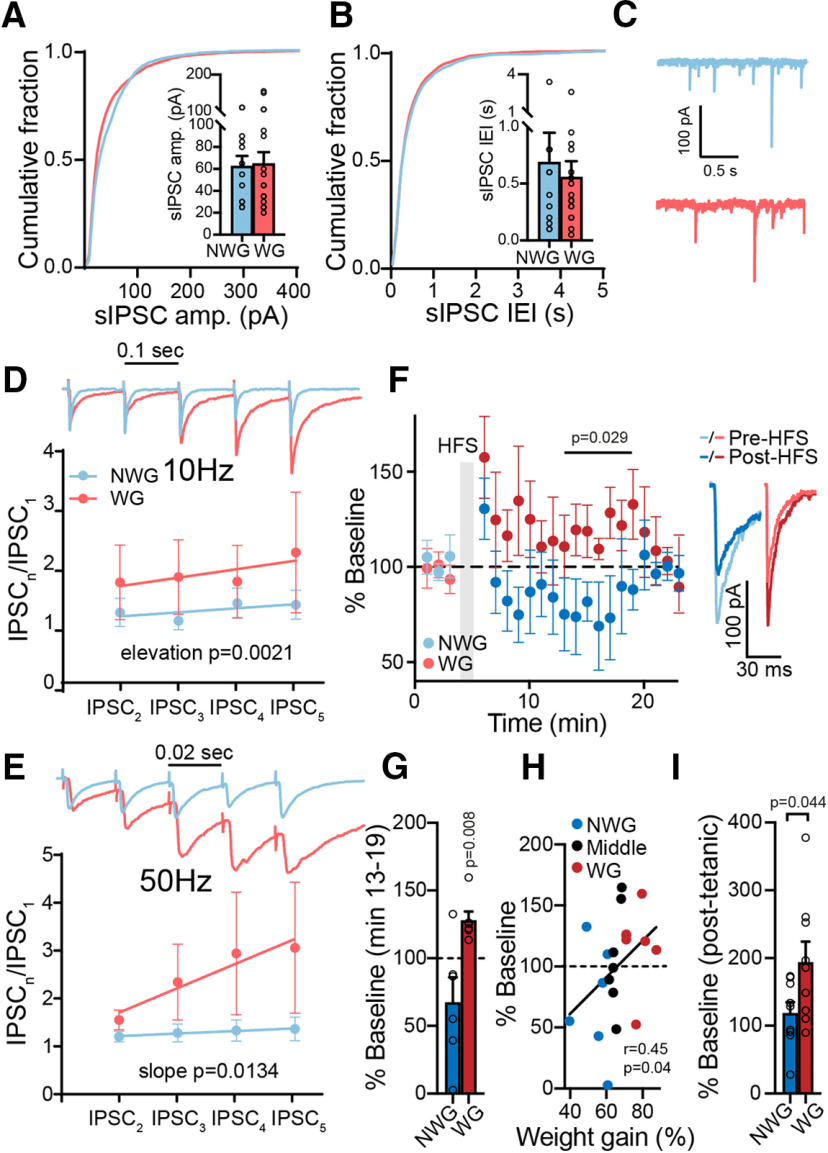
Figure 4.
Weight-gainer and non-weight gainer mice show different synaptic plasticities in their GABA input to the VP. A, B, sIPSC amplitude (amp.; A) and IEI (B) were similar between WG and NWG mice (Kolmogorov–Smirnov tests). Insets, Comparison of medians (unpaired two-tailed t tests). C, Representative sIPSC traces. D, E, Five consecutive pulses at 10 Hz (D) or 50 Hz (E) generated short-term potentiation of IPSCs in WG but not NWG mice (two-way ANCOVA tests). Insets, Representative traces synchronized with x-axis. Stimulation artifacts were truncated. F, G, HFS protocol given to inhibitory inputs to the VP generated a transient potentiation in WG mice but not in NWG mice (mixed-effects ANOVA; one-sample t test for comparing minutes 13–19, 100% of baseline). F, Inset, Representative traces. H, HFS-induced plasticity in VP neurons was positively correlated with weight gain (Middle, middle 33% weight gainers), going from depression in NWG mice to potentiation in WG mice. Correlation was assessed using nonparametric Spearman correlation. I, Post-tetanic potentiation, measured at the first time point after the HFS, was stronger in WG mice (unpaired two-tailed t test). Data are taken from 8–17 cells in 7 mice/group. Data are presented as the mean ± SEM.

