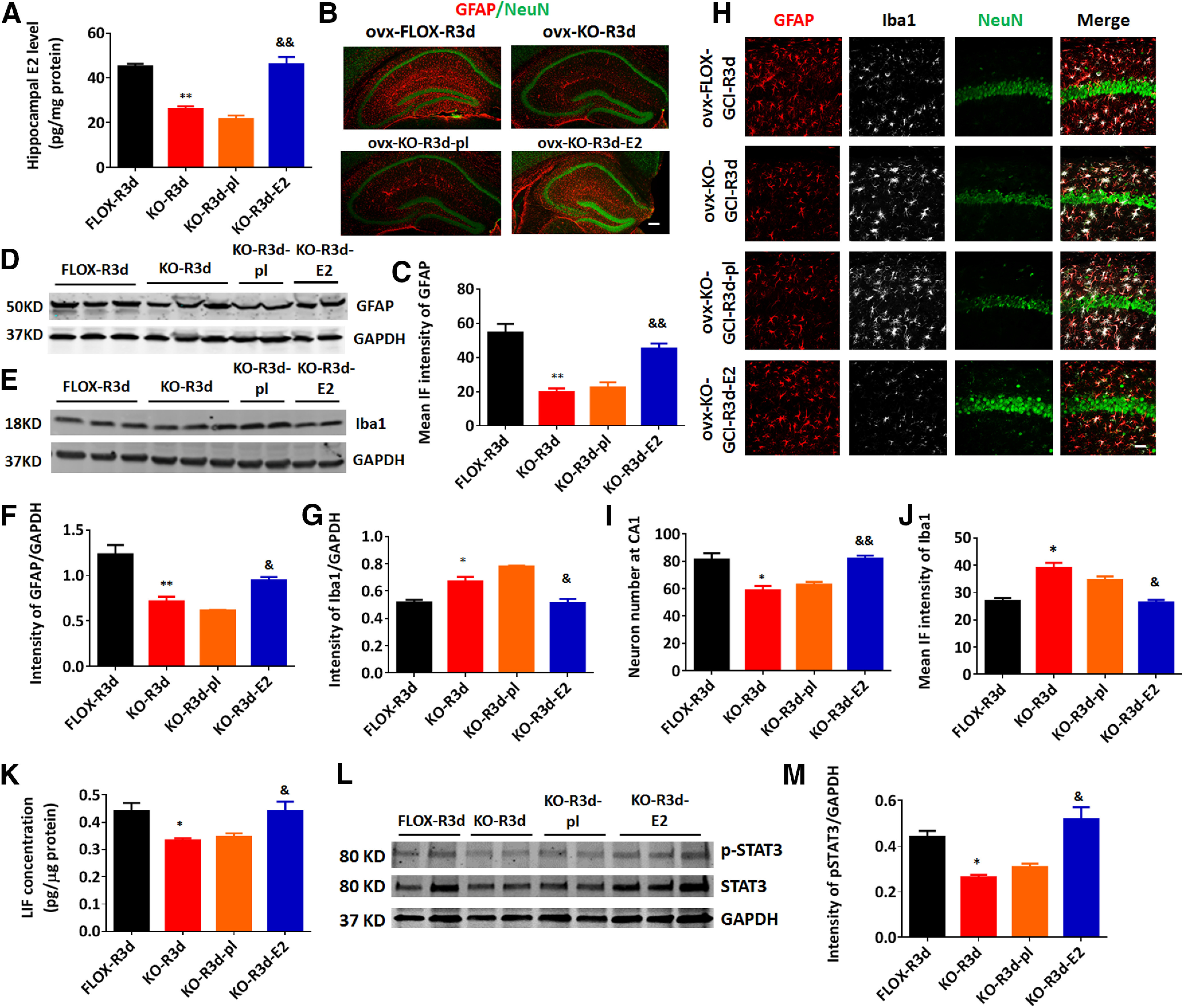
Figure 11.

Reinstatement of forebrain E2 levels by exogenous E2 administration rescues reactive astrogliosis, reverses the increase of microglia activation, and is neuroprotective in the ovx-female GFAP-ARO-KO mice. A, E2 levels in hippocampal tissue samples were measured by ELISA at R3d in ovx-female animals. B, C, Representative images of immunostaining for GFAP and NeuN (B) and quantification of the mean IF intensity of GFAP (C) in the ovx-female hippocampus. Scale bar: 50 μm. D–G, Western blot analysis and quantification of GFAP (D, F) and Iba1 (E, G) expression in hippocampal tissue samples collected from ovx-female animals. H–J, Representative images of immunostaining for GFAP, Iba1, and NeuN (H) in the hippocampal CA1 region of ovx-female mice, and quantification of the neuron number in the hippocampal CA1 region (I) and the mean IF intensity of Iba1 (J) in H. Scale bar: 20 μm. K, LIF levels in hippocampal tissue samples were measured by ELISA at R3d in ovx-female animals. L, M, Western blot analysis and quantification of p-STAT3 level in hippocampal tissue samples collected from ovx-female animals. Data are mean ± SEM; n = 6 biologically independent animals; *p < 0.05, **p < 0.01 compared with FLOX-R3d, two-way ANOVA; &p < 0.05, &&p < 0.01 compared with KO-R3d, one-way ANOVA followed by post hoc tests.
