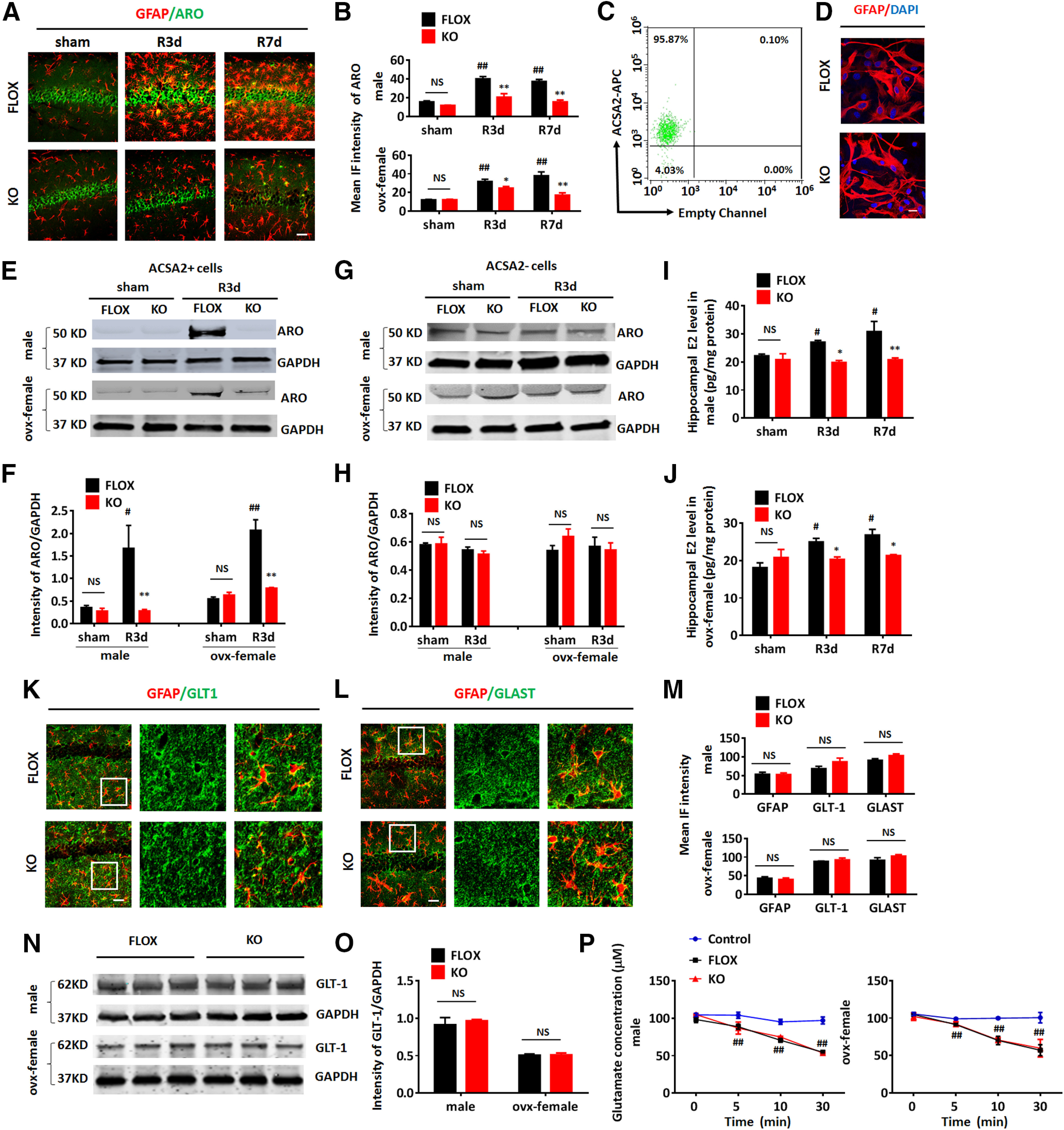
Figure 2.

Confirmation of aromatase deletion in astrocytes of GFAP-ARO-KO mice. A, Representative image of GFAP and aromatase (ARO) co-immunostaining in the hippocampal CA1 region of sham and reperfusion 3 and 7 d after GCI (R3d and R7d) of FLOX and GFAP-ARO-KO (KO) in male and ovx-female mice. Scale bar: 20 μm. B, Quantification of mean fluorescence intensity (IF) of aromatase in male and ovx-female hippocampal CA1. C, Flow cytometry analysis for ACSA2-APC in ACSA2+ cells isolated by magnetic cell sorting from the hemispheres of adult FLOX male mice. D, Immunocytochemistry demonstrates that purified adult astrocytes express GFAP at 24 h in vitro. Scale bar: 10 μm. E–H, Western blot analysis of aromatase on protein lysates of ACSA2+ astrocytes (E) and ACSA– cells (G) isolated from the hemispheres of male and ovx-female FLOX and KO mice in sham and R3d, and densitometry analysis (F, H) by ImageJ. Intensities of the aromatase band were normalized to the intensity of GAPDH from two independent experiments. Samples from three mice were pooled for one collection for each experiment. I, J, Hippocampal E2 levels were measured by ELISA assay on hippocampal tissue from sham, R3d, and R7d FLOX and KO mice in male (I) and ovx-female (J). K–L, Representative images of GFAP and GLT-1 (K) or GLAST (L) co-immunostaining in the hippocampal CA1 region of FLOX and KO mice under basal conditions. Scale bars: 20 μm. M, IF quantification of GFAP, GLT-1, and GLAST from IHC in male and ovx-female. N, O, Western blot analysis of FLOX and KO hippocampal tissue lysates from male and ovx-female and quantification of GLT-1 expression. P, Glutamate uptake analysis. Glutamate concentrations were measured after the glutamate containing media (100 μm) were incubated with FLOX/KO astrocytes isolated from male and ovx-female hemisphere for 0–30 min. Data are mean ± SEM; n = 4; #p < 0.05, ##p < 0.01 versus sham; *p < 0.05, **p < 0.01 versus KO with FLOX, two-way ANOVA followed by post hoc tests. NS, not significant.
