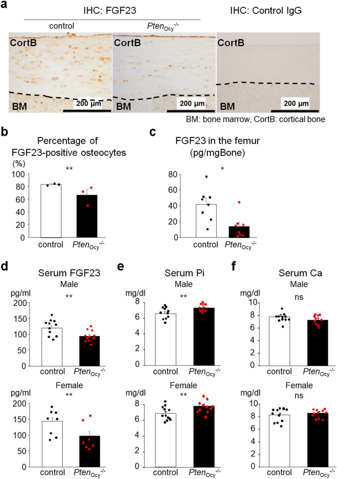
Figure 2.
Decreased intact FGF23 levels in PtenOcy−/− mice. (a) and (b) An immunohistochemical analysis of FGF23 in the femur was performed on 16-week-old male mice. A representative image of three independent experiments is shown (a). The percentage of FGF23-positive osteocytes over total osteocytes was counted (N = 3) (b). (c) The bone extracts of the femur were prepared from 16-week-old male mice and intact FGF23 concentration was measured (N = 8). (d–f) Serum levels of intact FGF23 (male: N = 11, female: N = 7–8) (d), Pi (male: N = 10–11, female: N = 11–13) (e), and Ca (male: N = 10–11, female: N = 11–13) (f) were measured in 16-week-old mice. Statistical analysis was performed by Mann–Whitney U test. *p < 0.01; **p < 0.05, ns: not significantly different.

