Figure 5.
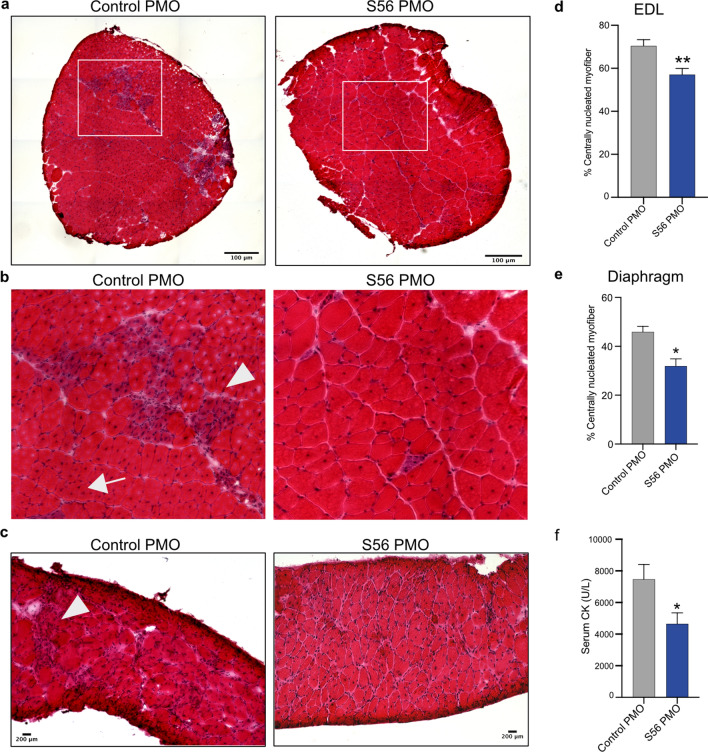
H&E staining of cryosections from control PMO or S56 PMO treated mdx mice EDL and diaphragm showed morphological improvement. (a) Representative H&E staining image of whole EDL cryosections (scale bar = 100 µm). (b) Regions highlighted by the white boxes were magnified (×3) (scale bar = 25 μm). (c) Representative H&E staining of diaphragm cryosections (scale bar = 200 µm). Control PMO treated EDL and diaphragm muscle cryosections show regenerated myofibers (arrow) and immune cell infiltration (arrowhead) and S56 PMO treatment appeared to alleviate symptoms. (d, e) Quantification of centrally nucleated EDL and diaphragm muscle fibers of mdx mice treated with control PMO or S56 PMO. The graphs show significant decrease in the percentage of CNFs of EDL and diaphragm muscles in S56 PMO treated mdx mice (**p = 0.002, n = 10 mice for both groups of EDL muscles, *p = 0.0140, n = 6 mice for both groups of diaphragm muscles). (f) The graph shows serum CK levels in S56 PMO treated mice were significantly lower than control PMO treated mdx mice (*p = 0.02, n = 10 mice for both groups). Each bar represents mean ± SEM. Statistical analysis performed by Mann–Whitney nonparametric test.

