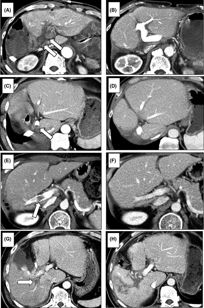
FIGURE 2.

CT findings of PVT. In patients with complete obstruction of MPV thrombosis—main grade 3 (CT on POD 1) (arrows) (A)—the thrombus was removed by urgent surgical thrombectomy (CT on POD 21) (B). In patients with a small MPV thrombosis—main grade 1 (CT on POD 9) (arrow) (C)—the thrombus resolved without treatment (CT at 6 mo after surgery) (D). In patients with right‐branch PVT—hilar thrombosis (CT on POD 7) (arrow) (E)—the thrombus resolved with anticoagulation therapy (CT at 3 mo after surgery) (F). In patients with posterior branch PVT—hilar thrombosis (CT on POD 10) (arrow) (G)—the thrombus organized in spite of anticoagulation therapy (CT at 1 mo after surgery) (H)
