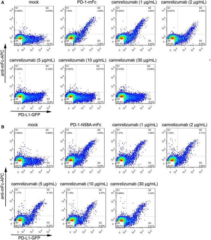
Figure 5. Reduced blocking efficiency of camrelizumab to N58 glycosylation‐deficient PD‐1.

-
A, BThe blocking of the binding of WT PD‐1-mFc (A) or N58A‐mutated PD-1‐mFc (B) to PD‐L1 transiently expressed on the surface of 293T cells are analyzed with varied concentrations of camrelizumab. The PD‐L1 expressing 293T cells stained with PBS alone is used as negative controls, whereas the cells stained with WT or N58A‐mutated PD‐1-mFc proteins are used as a positive control. The frequency of PD‐1-mFc staining positive cells in PD‐L1-GFP‐positive cells is labeled in the upright corner. The reduced frequency of the PD‐L1-mFc staining positive subpopulation compared with positive control indicates the blockage of the PD-1/PD-L1 interaction. The density of events at a given position in the plot is color‐coded, with red representing the highest number of events at that point in the graph, while yellow, green, and blue represent progressively lower event densities. The results presented here are representative of three independent experiments with similar results.
