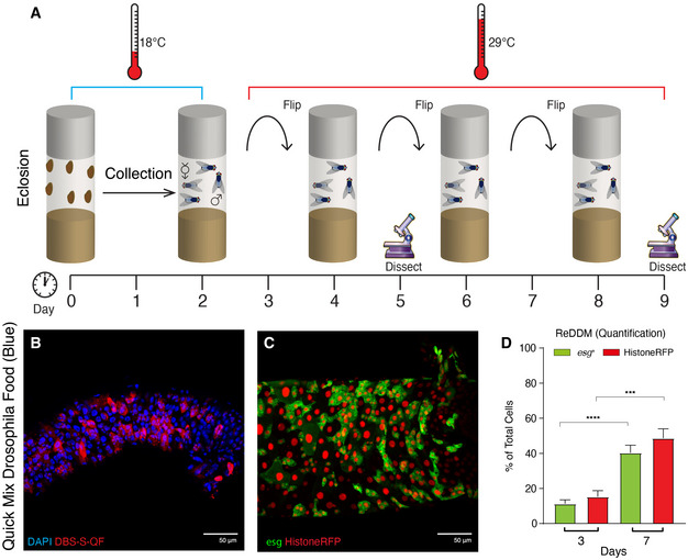
Figure EV1. Schematic of Drosophila experimental regime and results obtained using Drosophila Quick Mix Blue food.

- Parental Drosophila strains were crossed at 18°C and the progeny collected for 2 days post‐eclosion. After 2 days together, adult male and females flies were shifted at 29°C, where they were maintained and transferred to new vials with fresh food every two days. Experimental flies were dissected at 3 and/or 7 days post‐temperature shift at 29°C.
- Representative example of DBS‐S‐QF activation in (red, immunostaining anti‐HA) from flies reared in Drosophila Quick Mix Media (Blue) following the experimental regime described in A; note the widespread activation of the DBS‐S‐QF reporter 7 days post‐temperature shift at 29°C. DAPI (blue) labels the nuclei. Genotype: w1118 DBS‐S‐QF, UAS‐mCD8‐GFP, QUAS‐tomato‐HA.
- Representative example of the ReDDM labelling in a Drosophila intestine reared in Drosophila Quick Mix Media (Blue) following the experimental regime described in A; esg expression (green, GFP) labels intestinal progenitor cells (ISCs and EBs) and Histone‐RFP (red) acts as a semi‐permanent marker of differentiated intestinal cells as either EEs or ECs, after the esg promoter is silenced. Note the high number of Histone‐RFP‐positive cells without GFP signal as an indication of epithelial replenishment. Genotype: w1118; esg‐Gal4 UAS‐CD8‐GFP/Cyo; TubG80ts UAS‐Histone‐RFP
- Quantification of the ReDDM experiment shown in (C); note the significant increase of esg (****P < 0.0001) and Histone‐RFP (***P = 0.0004) labelled cells (Quantifications were made using N ≥ 2 biological replicates; unpaired two‐tailed t‐test, 3d n = 14, 7d n = 34). Error bars represent standard error of the mean. w1118; esg‐Gal4 UAS‐CD8‐GFP/Cyo; TubG80ts UAS‐Histone‐RFP.
