
Figure 4. Dronc is preferentially accumulated and activated in progenitor cells.
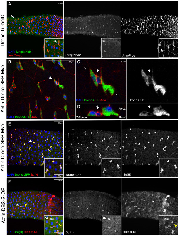
- Representative image of a Drosophila posterior midgut showing the expression of Armadillo (red membranes), Prospero (red nuclei of EEs) and Dronc‐TurboID (green shows Streptavidin‐labelled proteins by Dronc‐TurboID); note the preferential enrichment of Streptavidin staining is preferentially enriched in the intestinal progenitor cells (white arrows indicate an intestinal progenitor cell upregulating Dronc). Genotype: Dronc‐TurboID‐V5 (Masayuki Miura)/Tm6B.
- Representative image of a Drosophila posterior midgut showing the expression of Armadillo (red membranes) and Actin‐Dronc‐GFP‐Myc (green, anti‐GFP); note the preferential enrichment of Dronc expression in the small groups of cells formed by intestinal progenitor cells (arrow). Genotype: w 1118; Actin‐Dronc‐GFP‐Myc (attP‐VK37)/Cyo.
- High magnification of the picture shown in (B). The white arrow indicates a presumptive ISC showing low levels of Dronc compared with the flanking GFP + EBs. Genotype: w 1118; Actin‐Dronc‐GFP‐Myc (attP‐VK37)/Cyo. The arrow indicates a presumptive small ISC expressing high levels of Arm and low levels of Dronc‐GFP‐Myc.
- Transversal Z‐section of the cells shown in C. Genotype: w 1118; Actin‐Dronc‐GFP‐Myc (attP‐VK37)/Cyo. The arrow indicates a presumptive small ISC expressing high levels of Arm and low levels of Dronc‐GFP‐Myc.
- The enteroblast marker Su(H) (red, immunostaining against Beta‐galactosidase) strongly co‐localises with high levels of Dronc expression (green, immunostaining against GFP); white arrow indicates the enlarged area depicted in the insets. Genotype: w 1118, Su(H)GBE‐LacZ (Irene Miguel Aliaga); Actin‐Dronc‐GFP‐Myc (attP‐VK37)/Cyo.
- There is extensive overlap between the expression of the EB marker Su(H) (green, immunostaining against Beta‐galactosidase; white arrow) and the apical caspase reporter DBS‐S‐QF (red, immunostaining against HA), but it is also present in progenitor cells without Su(H) expression (yellow arrowheads). DAPI (Blue) labels cell nuclei in all the panels.
Data information: All of the experiments described in the figure apart from (F) were performed in Oxford medium following an experimental regime that protects the epithelial integrity at 29°C. The experiment shown in (F) was conducted at 25°C. Genotype: Females w 1118 Su(H)GBE‐LacZ/y1 w1118 UAS‐mCD8::GFP.L QUAS‐mtdTomato‐3xHA Act‐DBS‐S‐QF.