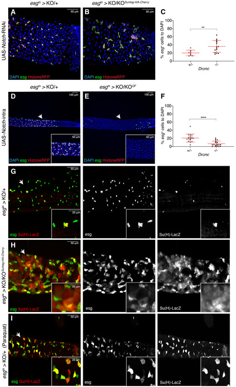
Figure 5. Dronc activation limits Notch signalling in the intestine.

- Representative ReDDM labelling of a Drosophila Dronc heterozygous (+/−) intestine overexpressing an RNAi against Notch for 3 days; note the lack of fully differentiated Histone‐RFP cells as EC (red) without esg expression (green, GFP). DAPI staining labels cell nuclei in panels A, B, D and E. All of the experiments described in the figure were performed in Oxford medium following an experimental regime that protects the epithelial integrity. Genotype: w 1118 UAS‐Notch‐RNAi (Joaquin Navascués); esg‐Gal4 UAS‐CD8‐GFP/+; TubG80ts UAS‐Histone‐RFP Dronc KO/+.
- Representative ReDDM labelling of a Drosophila Dronc mutant homozygous (−/−) intestine overexpressing an RNAi against Notch for 3 post‐temperature shift at 29°C; note the lack of differentiated Histone‐RFP cells as EC (red) without esg expression (green, GFP), as well as the increase of GFP‐positive cells (compare A to B). Genotype: w 1118 UAS‐Notch‐RNAi (Joaquin Navascués); esg‐Gal4 UAS‐CD8‐GFP/+; TubG80ts UAS‐Histone‐RFP Dronc KO/UAS‐Flippase (BL8209) FRT Dronc‐GFP‐APEX FRT suntag‐HA‐Cherry.
- Quantification of the number of Histone‐RFP cells normalised to DAPI (proxy of progenitor cell proliferation obtained from the experiments shown in A and B panels); note the statically significant increase in Histone‐RFP‐positive cells in Dronc homozygous mutant intestines compared with controls (**P = 0.0080) (Quantifications were made using N ≥ 2 biological replicates; unpaired two‐tailed t‐test, +/− n = 8, −/− n = 15). Error bars represent standard deviation of the mean. Genotypes: +/−: w 1118 UAS‐Notch‐RNAi; esg‐Gal4 UAS‐CD8‐GFP/+; TubG80ts UAS‐Histone‐RFP Dronc KO/+; −/−: w 1118 UAS‐Notch‐RNAi (Joaquin Navascués); esg‐Gal4 UAS‐CD8‐GFP/+; TubG80ts UAS‐Histone‐RFP Dronc KO/UAS‐Flippase (BL8209) FRT Dronc‐GFP‐APEX FRT suntag‐HA‐Cherry.
- Drosophila Dronc heterozygous intestine overexpressing the Notch intracellular domain for 7 days post‐temperature shift at 29°C; intestinal esg‐positive progenitor cells (green (GFP) and red (Histone‐RFP). Genotype: w 1118; esg‐Gal4 UAS‐CD8‐GFP/UAS‐Notchintra (Joaquin Navascués); TubG80ts UAS‐Histone‐RFP Dronc KO/+.
- Drosophila Dronc homozygous intestine overexpressing the Notch intracellular domain for 7d post‐temperature shift at 29°C; notice that the Dronc deficiency accelerates the elimination of intestinal progenitor cells induced by Notch overactivation (compare D and E). The white arrows indicate the position of insets 500 μm from the posterior region. Note the complete loss of esg‐labelled cells in this region. Genotype: w 1118; esg‐Gal4 UAS‐CD8‐GFP/ UAS‐Notchintra; TubG80ts UAS‐Histone‐RFP Dronc KO/UAS‐Flippase (BL8209) FRT Dronc‐GFP‐APEX FRT suntag‐HA‐Cherry.
- Relative number of esg‐positive cells to DAPI in either heterozygous or homozygous Dronc‐mutant esg cells overexpressing Notch intra; note the significant reduction of esg‐expressing cells (****P < 0.0001) (Quantifications were made using N ≥ 2 biological replicates; Mann–Whitney test, +/− n = 17, −/− n = 17). Error bars represent standard deviation of the mean. Genotypes: +/−: w 1118; esg‐Gal4 UAS‐CD8‐GFP/UAS‐Notchintra; TubG80ts UAS‐Histone‐RFP Dronc KO/+; −/−: w 1118; esg‐Gal4 UAS‐CD8‐GFP/UAS‐Notchintra; TubG80ts UAS‐Histone‐RFP Dronc KO/ UAS‐Flippase (BL8209) FRT Dronc‐GFP‐APEX FRT suntag‐HA‐Cherry.
- Representative image of a Drosophila Dronc heterozygous intestines 7 days after transfer to 29°C. esg expression in green (GFP) labels all intestinal progenitors cells, whilst Su(H)‐lacZ (red) distinguishes a subpopulation of EBs within the esg‐expressing cells. Genotype: w 1118, Su(H)GBE‐LacZ; esg‐Gal4 UAS‐CD8‐GFP/+; TubG80ts UAS‐Histone‐RFP Dronc KO/+.
- Representative image of a Drosophila Dronc KO intestine 7 days after transfer to 29°C. Note, the transcription of Su(H) in large cell GFP (−), suggestive of an aberrant upregulation of Notch signalling within fully differentiated ECs. Genotype: w 1118, Su(H)GBE‐LacZ; esg‐Gal4 UAS‐CD8‐GFP/+; TubG80ts UAS‐Histone‐RFP Dronc KO/UAS‐Flippase (BL8209) FRT Dronc‐GFP‐APEX FRT suntag‐HA‐Cherry.
- Representative image of a 7d Drosophila Dronc heterozygous intestines following a 16‐h treatment with paraquat. Note the absence of large Su(H) positive, GFP (−) cells compared with (H). The white arrows indicate the enlarged area depicted in the insets. Genotypes: +/−: w 1118, Su(H)GBE‐LacZ; esg‐Gal4 UAS‐CD8‐GFP/+; TubG80ts UAS‐Histone‐RFP Dronc KO/+; −/−: w 1118, Su(H)GBE‐LacZ; esg‐Gal4 UAS‐CD8‐GFP/+; TubG80ts UAS‐Histone‐RFP Dronc KO/UAS‐Flippase (BL8209) FRT Dronc‐GFP‐APEX FRT suntag‐HA‐Cherry.
Data information: (D–H) The white arrows indicate the enlarged area depicted in the insets.
