
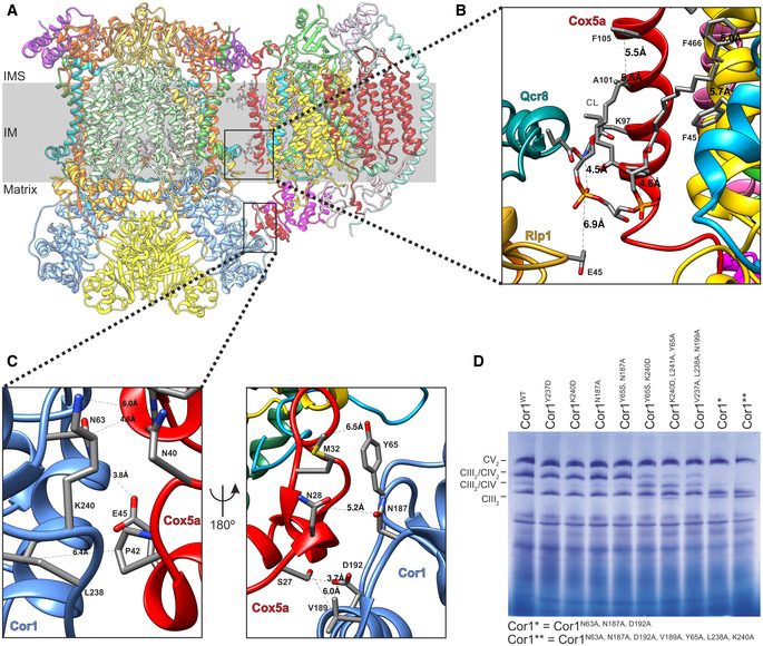
Figure 1. A detailed structure of the CIII‐CIV interface allows to design supercomplex‐disrupting mutations.
-
ASide view of the overall structure of the Saccharomyces cerevisiae CIII2/CIV supercomplex and its position in the mitochondrial inner membrane (IM). IMS: intermembrane space.
-
B, CZoom‐in of the CIII2/CIV interaction sites with residues and distances annotated. The inner membrane protein–lipid–protein interactions (Rip1‐CL-Cox5a), where Rip1 is marked in gold, cardiolipin (CL) in gray, and Cox5a in red (B), and the mitochondrial matrix, protein–protein interaction of Cor1 (blue), and Cox5a (red) (C) are shown.
-
DBlue‐native gel electrophoresis of S. cerevisiae strains with indicated mutations in Cor1.