
Figure 4. AMOTL1 promotes Merlin ubiquitination by mediating the interaction between NEDD4L and Merlin.
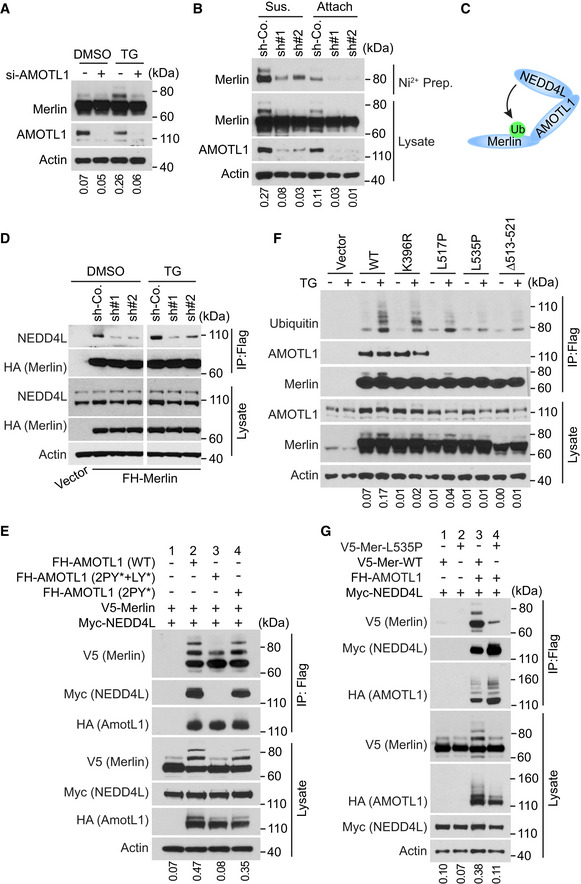
- LN229 cells transduced with a pool of four siRNAs against AMOTL1 (+) or a scrambled siRNA (−) were treated with DMSO or thapsigargin (TG) and subjected to Western blotting. The ratio of mono‐ubiquitinated to native Merlin in each lane was quantified by ImageJ and is shown under the blot.
- LN229 cells stably transduced with 6×‐Histidine‐tagged Ubiquitin (His‐Ubi) were then stably transduced with indicated shRNAs against AMOTL1 or a scrambled shRNA control (sh‐Co.). These cells were detached, lysed, and subjected to nickel‐charged affinity purification followed by Western blotting for endogenous Merlin. The ratio of mono‐ubiquitinated to native Merlin in each lane of the lysate blot was quantified by ImageJ and is shown under the blot.
- A diagram illustrating that AMOTL1 mediates the NEDD4L‐Merlin interaction.
- Merlin‐depleted LN229 cells stably expressing Flag‐HA-tagged Merlin were transduced with scrambled shRNA (sh‐Co.) or two distinct shRNAs targeting AMOTL1, treated with DMSO or thapsigargin (TG) and subjected to Flag immunoprecipitation followed by Western blotting.
- HEK293T cells were transfected with the indicated constructs. The cells were lysed and subjected to immunoprecipitation with a Flag antibody. The lysate and immunoprecipitated products were subjected to Western blotting. LY, LPTY motif; PY, PPEY motif. The ratio of mono‐ubiquitinated to native Merlin in each lane of the lysate blot was quantified by ImageJ and is shown under the blot.
- Merlin‐depleted LN229 cells stably transduced with Flag‐tagged wild‐type Merlin or the indicated mutants were treated with DMSO or thapsigargin (TG). The cells were lysed and subjected to immunoprecipitation with a Flag antibody. The lysate and immunoprecipitated products were subjected to Western blotting. The ratio of mono‐ubiquitinated to native Merlin in each lane of the lysate blot was quantified by ImageJ and is shown under the blot.
- HEK293T cells were transfected with the indicated constructs. The cells were lysed and subjected to immunoprecipitation with a Flag antibody. The lysate and immunoprecipitated products were subjected to Western blotting. The ratio of mono‐ubiquitinated to native Merlin in each lane of the lysate blot was quantified by ImageJ and is shown under the blot.
Source data are available online for this figure.