Abstract

Editorial expression of concern
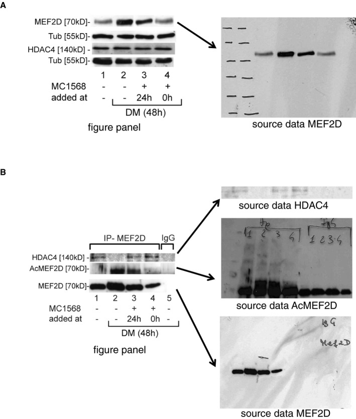
We were alerted to apparent image aberrations in Fig 2B of this paper, and subsequently identified further inconsistencies in Fig 2B and C. Figure 2B, panel MEF2D, shows a striking similarity between the first and fourth band, and a potential splice line between band 3 and band 4. A digital image provided by the authors as representative of the source data for lane MEF2D of Fig 2B (see Image A below) does not match to the published image in one of the four lanes (lane 3). The authors did not provide an explanation for this discrepancy. Figure 2B is suggested to show that the class II‐specific HDAC inhibitor MC1568 arrests myogenesis by decreasing the levels of the myocyte enhancer factor 2D (MEF2D). The authors describe that the panel shows levels of MEF2D upon differentiation induction and parallel treatment with the HDAC inhibitor. MEF2D is suggested to be induced during differentiation (lane 2—48 h of differentiation medium), compared to lane 1 (control). This differentiation‐specific MEF2D induction is suggested to be reduced by inhibitor treatment (lane 3 and lane 4). These conclusions are central to the paper.
In Fig 2C, panel HDAC4, we observed similar background patterns in lanes 2 and 5. Panel AcMEF2D contains inconsistent background patterns in lane 5 (IgG—negative IP control), compared to the rest of the panel, consistent with splicing. In the panel MEF2D, lane 5 (IgG—negative IP control) does not show any signal, suggesting that the original image data of this lane are missing. The authors provided source data for this experiment, which they claim represents unmodified images of the data underlying the figure panels in 2C (see Image B below). These are consistent with a lack of a specific signal in the IgG lanes in these experiments. However, it remains unclear if the source data provided unequivocally and completely represent the displayed figure.
Figure 2C is suggested to show that the HDAC‐MEF2 complex is stabilized by inhibitor treatment by a co‐immunoprecipitation experiment. In undifferentiated cells, HDAC4 is suggested to co‐precipitate with ac‐MEF2D (lane 1). After 48 h of differentiation, it does not (lane 2). However, when the inhibitor is added, both proteins are suggested to co‐immunoprecipitate, also upon differentiation induction (lanes 3 and 4, respectively).
Both panels concern significant aspects of the scientific conclusions of the manuscript, in particular that class II HDAC inhibitors impair myogenesis by modulating the stability and activity of HDAC‐MEF2 complexes.

We note that the authors provided source data for replicate experiments that they state were performed at the same time as the published data for both panels (consistent with the labelling on the original films). These data are publicly available on FigShare (https://figshare.com/articles/Raw_data_from_Nebbioso_et_al_EMBO_Rep_2009_10_776-82/12349841). The authors’ research institution also provided figures that are stated to represent replicate experiments similar to the ones shown in Fig 2B and 2C of the original publication performed as part of their investigation into these issues by independent investigators using reagents and methods provided by the authors.
Further, we have been informed by Antimo Migliaccio, head of the department which corresponding author Lucia Altucci is presently affiliated to Dipartimento di Medicina di Precisione, Università degli Studi della Campania "Luigi Vanvitelli", Napoli, that an institutional investigation formally concluded that no illicit image manipulation is apparent in the manuscript.
In line with EMBO Press’s transparent publishing policy, the Appendix includes key communication between journal, authors and research institution. The Appendix also includes the image integrity analyses by the journal as well as the institution.
Author statement
All of the authors state that the conclusions drawn from figure 2B and 2C, and from the paper as a whole remain unchanged. All authors agree with the publication of the corrigendum in this form.
Panel A – Originally published version of Fig 2, panel B (left), and the source data as provided by the authors (right).
Panel B – Originally published version of Fig 2, panel 2C (left) and the source data as provided by the authors (right).
Supporting information
Appendix
Correction to: EMBO Reports (2009) 10: 776–782. DOI 10.1038/embor.2009.88 ¦ Published online 5 June 2009
Associated Data
This section collects any data citations, data availability statements, or supplementary materials included in this article.
Supplementary Materials
Appendix


