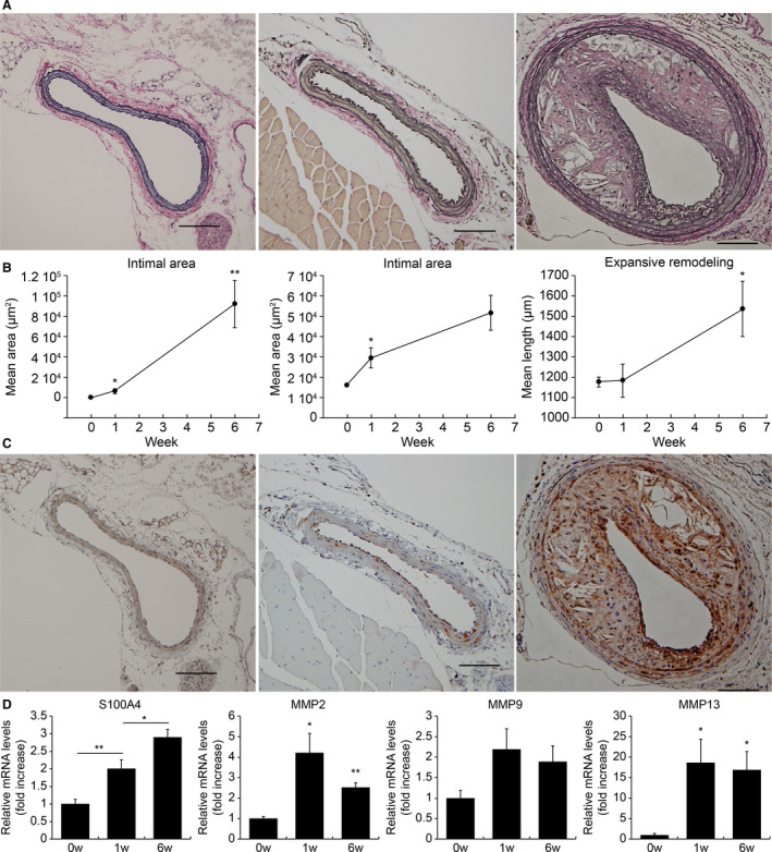
Figure 3. Expression of S100A4 (S100 calcium‐binding protein A4) in the plaques of a mouse partial carotid ligation model.

A, Mouse left carotid arteries (CAs) stained with Elastica van Gieson at 0, 1, and 6 weeks after partial ligation. Representative images are as follows: e left=0 weeks, center=1 week), and right=6 weeks. Atherosclerotic lesions of the left CAs at 6 weeks after ligation exhibited expansive arterial remodeling with the proliferation of both intimal and medial lesions. B, Intimal area, medial area, and perimeter of the external elastic lamina of partially ligated CAs. For each group, the average values of 5 mice were calculated, and changes over time were evaluated. The intimal area had already expanded 1 week after ligation and then expanded further after 6 weeks. The medial area also showed a significant increase at 1 week after ligation and showed a tendency to further expand at 6 weeks after ligation. Expansive remodeling was observed at 6 weeks after ligation. C, Mouse left CAs stained with anti‐S100A4 antibody at 0, 1, and 6 weeks after partial ligation. Representative images are as follows: left=0 weeks, center=1 week, and right=6 weeks. S100A4 was expressed in the medial area at 1 week after ligation, and extensive expression was observed in both the medial and neointimal areas 6 weeks after ligation. D, The expression levels of S100a4, Mmp2, Mmp9, and Mmp13 at 0, 1, and 6 weeks after ligation (n=6–9 each time point). The expression of S100a4 showed a statistically significant increase over time. The expression levels of Mmp2 and Mmp13 increased from 1 week after ligation and showed a significant increase even after 6 weeks. In contrast, there was no significant difference in the expression levels of Mmp9. The gene expression levels were evaluated using quantitative PCR. *P<0.05, **P<0.01 for the indicated comparisons. MMP indicates matrix metalloproteinase.
