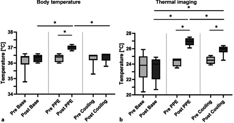
Fig. 2.
Results of the body temperature (BT) measurements and thermal imaging. a Body temperature. b Thermal imaging. BT measurements and thermal imaging were performed before and after the activity period. In contrast to pre-cycle levels, a significant increase of BT (p = 0.031) and surface temperature (p = 0.031) was shown post-PPE cycle. Boxplots represent median, quartiles and min–max values. An asterisk indicates statistical significance

