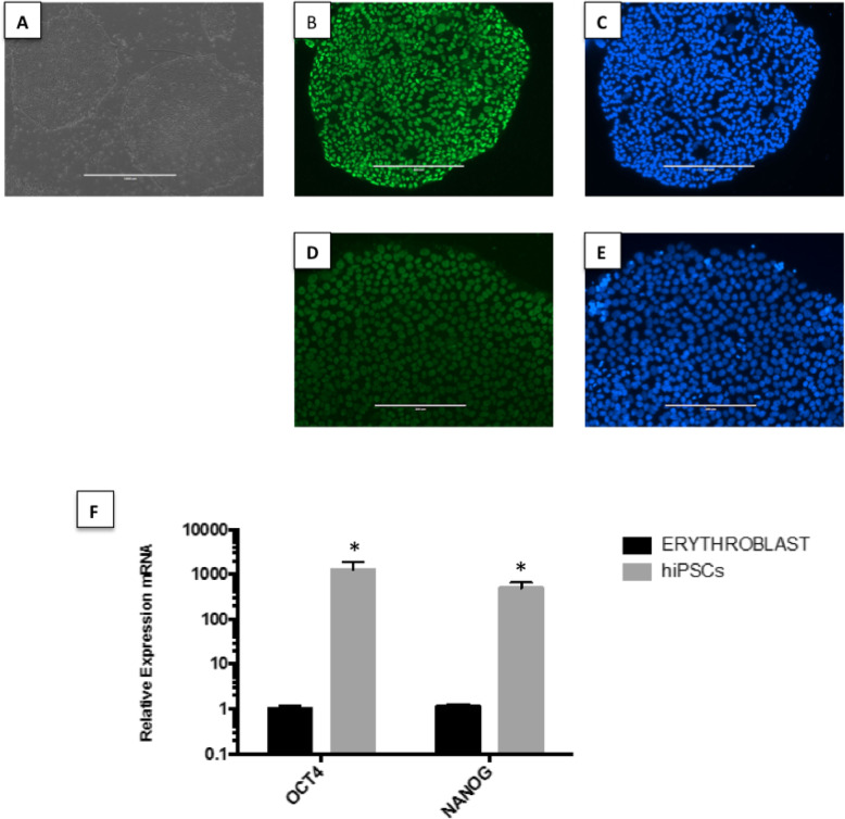
Fig. 1.
Characterization of peripheral blood-derived hiPSCs. a Typical morphology of undifferentiated hiPSC colonies. The colonies grown over Matrigel have a homogeneous shape, smooth and regular edges, and positive staining for b OCT4, d NANOG, and c, e DAPI staining for the same colonies of b and d, respectively. Scale bar 200 μm. f Gene expression levels in samples collected before (erythroblasts) and after (hiPSCs) reprogramming, generated by referencing each gene to HMBS expression levels as an internal control. Healthy erythroblast mRNA was used as the comparative sample. *p value < 0.05 vs. erythroblast

