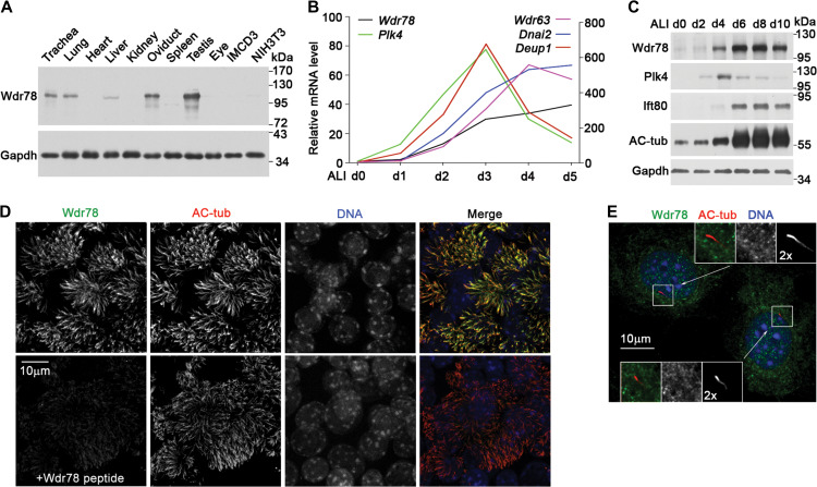
Figure 2.
Wdr78 is located in motile cilia. (A) Wdr78 was highly expressed in motile cilia-containing tissues, including trachea, lung, oviduct, and testis. Cultured IMCD3 and NIH3T3 cells were serum-starved for 48 h to induce primary cilia. Gapdh served as loading control. (B) Expression profile of Wdr78 during multiciliogenesis. qRT-PCR was performed using mRNAs extracted from mTECs cultured at an ALI for the indicated days (d). Plk4 and Deup1 served as markers for centriole amplification, whereas Wdr63 and Dnai2 served as markers for multiciliogenesis. The results were representative of two independent sets of experiments. (C) Protein levels of Wdr78 during the multiciliogenesis of mTECs. The increased levels of Ift80 and acetylated tubulin (AC-tub) indicate multiciliogenesis. Plk4 and Gapdh served as marker for centriole amplification and loading control, respectively. (D) Wdr78 localized in the axonemes of mTEC cilia. mTECs were fixed at ALI d7 and stained with anti-Wdr78 antibody preincubated with or without the Wdr78 peptide that was used for antibody generation. AC-tub marks ciliary axonemes. Nuclear DNA was stained with DAPI. (E) Wdr78 was not a primary ciliary protein. NIH3T3 cells were serum-starved for 48 h to induce primary ciliary formation. AC-tub marks ciliary axonemes. Nuclear DNA was stained with DAPI.

