Figure 5.
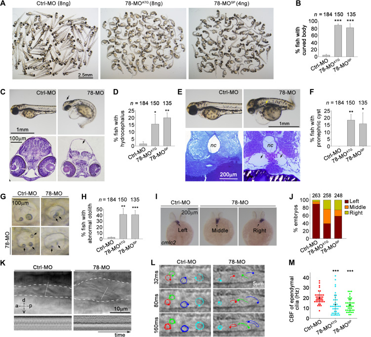
Phenotypes of zebrafish Wdr78 morphants. (A and B) Body curvatures at 72 hpf. Ctrl-MO, the standard control morpholino; 78-MOATG and 78-MOsp, two antisense MOs targeting the translation initiation site and a splicing site of Wdr78 mRNA, respectively. The same populations were also used for assays in C–H. (C and D) Hydrocephalus (arrows). (E and F) Pronephric cysts (arrows). nc, notochore. (G and H) Abnormal number, size, or positioning of otoliths (arrows). (I and J) Randomized heart tube orientations. Whole-mount in situ hybridization of Cmlc2 mRNA was performed to label the heart tube (arrows) at 24–27 hpf. (K) Impaired motility of pronephric cilia. Motility of pronephric cilia, recorded at 250 frames per sec (fps) as differential-interference-contrast images at 60 hpf, is presented in Supplementary Videos S4 and S5. The first frames of the videos, with the pronephric duct outlined with dashed lines and kymographs of the cilia at the position of the white lines are shown here. a, anterior; d, dorsal; p, posterior; v, ventral. (L and M) Impaired motility of spinal cord cilia. Motilities of spinal cord cilia were recorded at 250 fps at 60 hpf. Trajectories of four cilia during the first 160 ms of imaging are shown for each embryo. Please refer to Supplementary Videos S6 and S7. The CBFs of three traceable cilia were quantified for each of 13 morphants in each group. Note that immobile cilia, if any, were difficult to identify in the images due to high backgrounds. Quantification results were from three independent experiments and presented as mean ± SD. Embryo numbers analyzed are listed over each histogram. Student’s t-test: *P < 0.05; **P < 0.01; ***P < 0.001.

