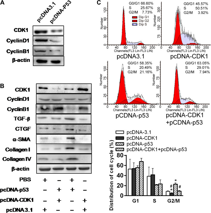
Figure 5.
p53 suppresses expression of CDK1 and profibrotic proteins in HK-2 cells. (A) Western blotting analyses of CDK1, Cyclin D1, and Cyclin B1 expression in p53 overexpression plasmid or control vector-treated HK-2 cells under normoxia. (B) Western blotting analysis of protein expression in p53 and/or CDK1 overexpression plasmid-treated HK-2 cells under normoxia. Blots shown are representative of three independent experiments. (C) Cell cycle distribution in HK-2 cells transfected with pcDNA3.1 empty vector, pcDNA-CKD1, or/and pcDNA-p53 under normoxia was analyzed by flow cytometry (left). Changes in the percentage of proliferating cells in G2/M phase are shown (right).*P < 0.05 vs. empty vector-transfected cells; #P < 0.05 vs. pcDNA-p53-transfected cells.

