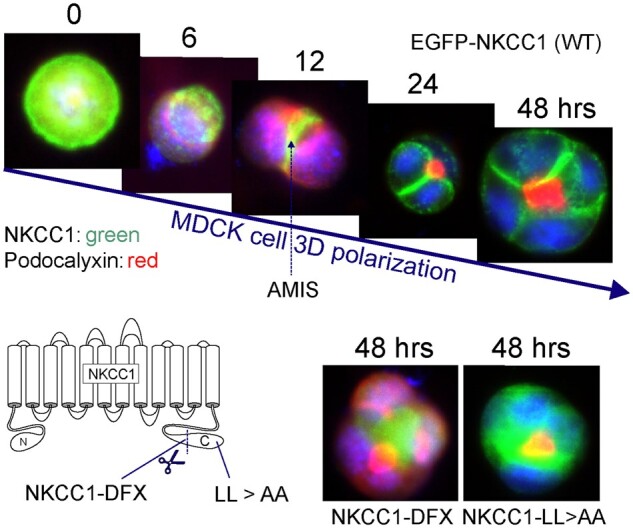
Figure 1.

Membrane Localization of NKCC1 During Polarization of MDCK Cells in Matrigel. Top panels: MDCKII cells were transfected with EGFP-tagged NKCC1 and plated in Matrigel. As a single cell divide to create a polarized structure, NKCC1 which is first seen all around the cell now concentrates at the Apical Membrane Initiation Site prior to localizing at its final destination: the basolateral membrane. The process takes 24–48 h to create the first four cell polarized structure. Bottom panels: This process is prevented by truncating the carboxyl-terminus of NKCC1, as in the NKCC1-DFX patient or by mutating a dileucine motif located at the extreme carboxyl-terminal tail of the cotransporter. In each case, NKCC1 staining colocalizes with podocalyxin at the apical pole. NKCC1 is labeled in green; whereas the apical marker podocalyxin is labeled in red. Data were redrawn from Koumangoye et al.16
