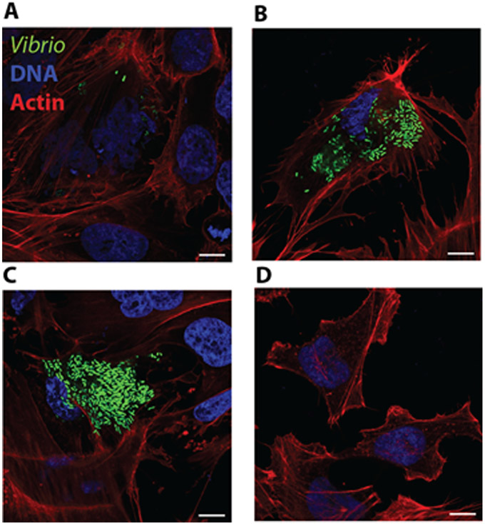
Fig 5.
Confocal micrographs of HeLa cells invaded by V. parahaemolyticus GFP expressing CAB2 strain at A)1, B)3, C)5 and D)7h post gentamicin treatment during a gentamicin protection assay.
HeLa cell actin was stained with rhodamine phalloidin (red) and DNA was stained with Hoechst (blue). Scale bars = 10 μm.

