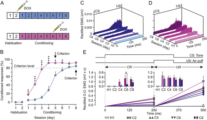
Figure 8.

Classical conditioning of eyelid responses during the inhibition of both CLs. CL neurons were inhibited by the local injection of a cocktail of rAAVs equipped with doxycycline (Dox)-dependent tetracycline-controlled genetic switches, which release tetanus toxin (TeTxLC) when activated. (A) Animals (n = 8) were classically conditioned using a delay paradigm following two protocols: half of them (blue group) were injected with Dox after the second habituation session and the other half (magenta group) after the sixth conditioning session. (B) Learning curves corresponding to the two groups of animals. Data are presented as mean ± SEM (see the multiple comparison reports: *P < 0.05; **P < 0.01; ***P < 0.001; Holm–Sidak or Tukey–Kramer tests). (C) Rectified EMG activity of the left O.O. muscle from the blue group during the CS–US interval (CRs time gap) and collected for the indicated habituation and conditioning sessions (n = 50 trials per session from n = 4 animals). (D) Same representation for magenta group. Note the earlier and larger CRs attained by this group during CS–US interval. (E) Quantitative analysis of cumulative areas (in mV × ms) of the rectified EMG activity of the left O.O. muscle recorded during the CS–US interval (CR time gap) and during the 250 ms following it (CS + US interval plus 150 ms, UR time gap) for the five indicated sessions. The insets illustrate the differences in net EMG areas between the two groups (*P < 0.05) during the CS–US (CRs) and the CS + US intervals (URs). No differences were found between groups for URs.
