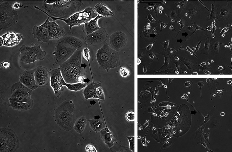
Figure 2.
Representative examples of TNTs (indicated along their length by arrowheads) connecting ovarian carcinoma cells in culture. The images on the left are of cells from the OvCar 3 cell line. The two images on the right are of cells derived from a malignant effusion (ascites) fluid from women with advanced ovarian malignancy. Scale bars = 100 μm.

