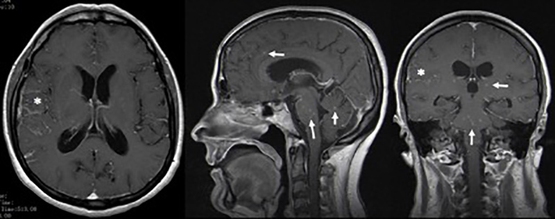
Figure 1.

Cerebral magnetic resonance imaging demonstrating contrast enhanced small intra-axial lesions in the supratentorial and infratentorial compartments with a miliary pattern (-->), and leptomeningeal enhancement of basal cisterns, extending to the opercular region on the right (*).
