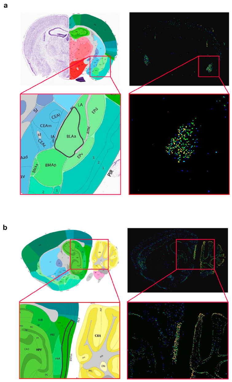
Figure 2.

Localized Wwox expression in basolateral amygdala and medial entorhinal cortex of mouse brain. (a) Coronal brain section showing Wwox mRNA in-situ hybridization signals clearly delineating the anterior (BLAa) region of the basolateral amigdalar nucleus in the cortical subplate. The inset shows the zoomed image of the BLAa region, highlighted with a black border in the annotated panel (left). (b) Sagittal section showing in situ hybridization signals, specifically lighting-up layer 2 of the medial entorhinal cortex (ENTm2, medial part, dorsal zone, layer 2). The inset shows the zoomed image of the ENTm2 region, highlighted with a black border in the annotated panel (left). It can also be observed that Wwox expression clearly delineates specific layers of the cerebellar cortex. Brain tissue sections from 56-day old, C57Bl/6J male mouse. All images were obtained from the Allen Mouse Brain Atlas—Allen Institute (https://mouse.brain-map.org/) [19].
