Figure 6.
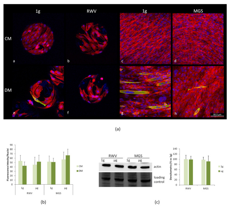
C2C12 were cultured in the RWV and in the MGS or in 1 g-ground control for 4 days. (a) The cells were fixed and immunofluorescence was performed using antibodies against myosin heavy chain 3 (MHC). The samples were counterstained with rhodamine labeled phalloidin and 4′,6-diamidine-2′-phenylindole dihydrochloride (DAPI). (b) Phalloidin fluorescence was quantified by ImageJ. (c) Cell extracts were analyzed for β-actin levels by western blot, and densitometry was performed using ImageJ.

