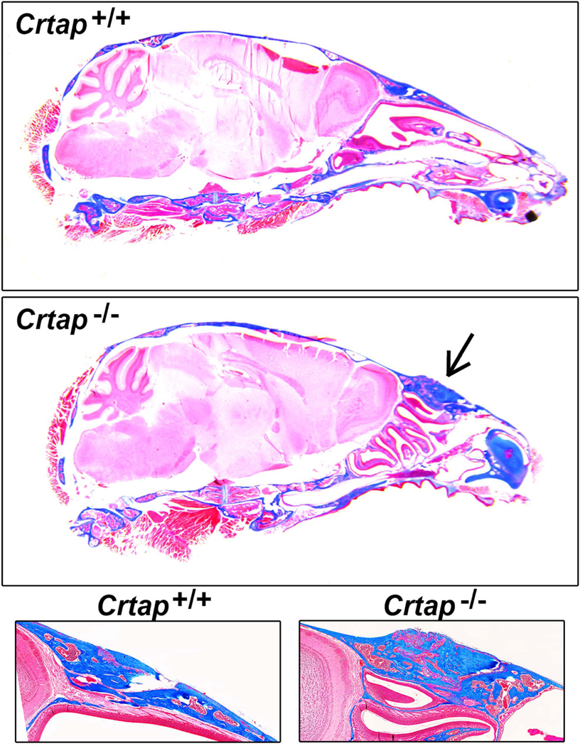
Figure 2. Crtap−/− Mice Exhibit Fusion of Frontal and Nasal Bones.

Histologic Masson’s trichrome stain on decalcified tissue sections reveals thickened and dysmorphic anterior frontal and posterior nasal bone in addition to fusion of the nasofrontal suture (black arrow). These defects were seen in all Crtap−/− mice (5/5) but no Crtap+/+ mice at 2 months.
