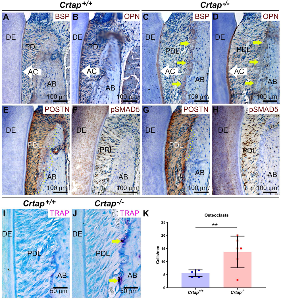
Figure 6. Altered Periodontal Markers in in Crtap−/− Mice.

Immunohistochemistry reveals altered (A, C) bone sialoprotein (BSP) and (B, D) osteopontin (OPN) in Crap−/− vs. control alveolar bone (AB) adjacent to periodontal ligament (PDL) (indicated by yellow arrows), as well as a thicker acellular cementum (AC) layer. DE=dentin. (E, G) While periostin (POSTN) localizes to PDL and embedded Sharpey’s fibers (indicated by yellow dotted line) in AB of control mice, reduced numbers of POSTN-positive Sharpey’s fibers are included in Crtap−/− AB. (F, H) Increased numbers of cells positive for phosphorylated SMAD5 (pSMAD5) are present in the PDL of Crtap−/− vs. control mice. (I, J) Increased numbers of tartrate-resistant acid phosphatase positive (TRAP+) osteoclast-like cells are observed on Crtap−/− vs. control AB surfaces. (K) Quantification reveals more than 2-fold greater numbers of osteoclast-like cells in Crtap−/− vs. control mice (**p<0.01).
