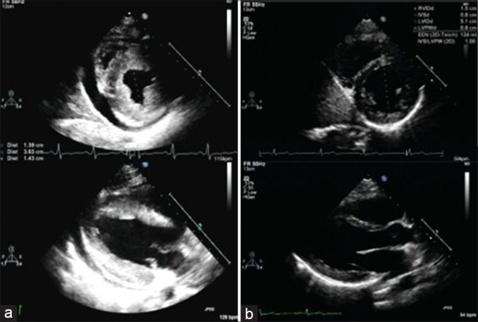
Figure 2.

(a) Initial short- and long-axis views demonstrating echo bright and hypertrophied heart, with a septal thickness of 14 mm (b) 4 months after presentation, the same views demonstrating complete resolution of hypertrophy, with septal thickness normalized to 8 mm
