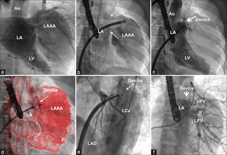
Figure 3.
Catheter closure of ostium of left atrial appendage aneurysm. Still-frame of rotational left atrial angiogram (a) identified right anterior oblique projection 26° degree as the best imaging plane [Online Video] to separate the left atrium and the aneurysm (b) that helped deployment of a 24-mm postmyocardial infarction muscular ventricular septal occluder device (c) using overlay imaging (d). Absence of left circumflex artery compression (e) and left upper pulmonary vein and lower pulmonary vein impingement (f) by the device was confirmed. LAD: Left anterior descending interventricular artery

