
Figure 4.
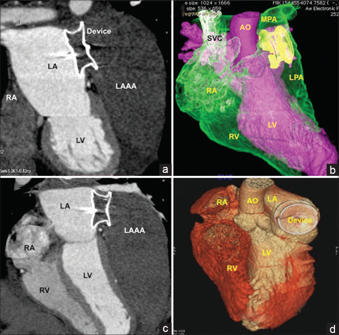
Complete thrombosis of the aneurysmal sac. Immediate postprocedural computed tomography in multiplanar reformatted (a and c) and volume-rendered images (b and d) showing complete closure of the ostium of the aneurysm leading to thrombosis in the entire sac. The left atrial disc of the device was well opposed to left atrial walls to promote future endothelialization